In the rapidly evolving landscape of oncology, precision medicine continues to drive transformative advances in cancer treatment and prognostication. A groundbreaking study recently published in BMC Cancer introduces an innovative multi-channel deep learning radiomics model designed to predict postoperative overall survival (OS) in patients diagnosed with laryngeal carcinoma. Leveraging contrast-enhanced computed tomography (CECT) images, this model not only enhances risk stratification but also holds promise for guiding individualized therapeutic decisions.
Laryngeal carcinoma, a malignancy affecting the voice box, presents unique challenges due to its anatomical complexity and functional significance. Prognosis after surgical intervention depends heavily on multiple patient-specific and tumor-related factors. Traditional staging systems, while useful, often fall short in capturing the full heterogeneity of tumor biology and patient response. Consequently, there is a crucial need for more sophisticated and quantitative methods that integrate imaging and computational analysis to refine survival predictions.
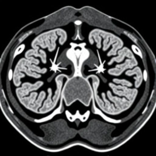
This study harnessed the power of radiomics—a discipline that translates medical images into high-dimensional data—and incorporated deep learning techniques to extract meaningful patterns from preoperative CECT scans. The researchers retrospectively gathered data from 272 individuals treated between 2016 and 2021 across two separate medical centers, ensuring the robustness of their findings through an external validation cohort.
The investigative team constructed two distinct imaging signatures. The first characterized traditional radiomics features encoding phenotypic expressions of the tumor microenvironment, while the second capitalized on multi-channel deep learning networks capable of discerning complex hierarchical imaging patterns beyond human perception. These signatures were generated from venous-phase images, highlighting contrast enhancements that reflect vascular and structural tumor characteristics.
A meticulous feature selection pipeline was employed, incorporating reproducibility assessments to guarantee stability, Spearman correlation to minimize redundancy, and least absolute shrinkage and selection operator (LASSO) regression to identify the most prognostically relevant variables. This subtle balance between interpretability and complexity is key to developing clinically feasible models without overfitting.
To maximize predictive accuracy, the study deployed ten distinct machine learning algorithms across the imaging signatures. Each algorithm offered unique strengths in pattern recognition and classification, and their performances were rigorously compared. These signatures formed the foundation for an integrated Deep Learning Radiomics Nomogram (DLRN), which seamlessly combined quantitative imaging biomarkers with clinical parameters to output individualized survival probabilities.
Evaluation metrics affirm the model’s robust prognostic capabilities. In the external test set, the DLRN achieved area under the curve (AUC) values of 0.74, 0.75, and 0.80 at 1-, 2-, and 3-year postoperative intervals respectively, alongside a Harrell’s concordance index (C-index) of 0.73. These metrics demonstrate superior discrimination compared to models relying solely on radiomics or deep learning features, underscoring the complementary power of a multi-channel strategy.
Critical to clinical adoption is model calibration and net benefit assessment. Calibration curves revealed excellent agreement between predicted and observed survival, instilling confidence in practical utility. Decision curve analysis (DCA), which weighs clinical decisions’ benefits against potential harms, confirmed the DLRN’s highest net benefit across relevant threshold probabilities, signaling strong potential to influence patient management.
Beyond aggregate performance, subgroup analyses illuminated the model’s consistency across diverse patient categories, including various clinical stages, age brackets, and surgical modalities. This generalizability suggests that the DLRN can serve a broad spectrum of laryngeal cancer cases, addressing the heterogeneity that often hampers one-size-fits-all prognostic tools.
The implications of this study are profound. By integrating advanced imaging analytics with deep learning, oncologists may soon be equipped with non-invasive, precise instruments to stratify risk at an individual level, tailoring postoperative surveillance and adjuvant therapy accordingly. Ultimately, this personalized approach has the potential to improve survival rates and optimize resource allocation in head and neck oncology.
Technically speaking, the fusion of radiomics and deep learning exploits both handcrafted and automatically derived features, capturing nuanced tumor characteristics. While radiomics traditionally relies on predefined image descriptors such as texture, shape, and intensity, deep learning algorithms learn hierarchical features directly from raw pixel data, offering complementary insights. The multi-channel architecture facilitates simultaneous processing of these distinct feature types, increasing predictive robustness.
The retrospective design, while extensive, accentuates the need for prospective validation in larger cohorts and multi-institutional settings to confirm clinical efficacy and reproducibility. Furthermore, integration with molecular and genomic markers, currently unexplored in this model, could enhance its predictive accuracy and unveil biological underpinnings of imaging phenotypes.
Looking forward, the study opens avenues for extending similar multi-modal deep learning frameworks to other tumor sites and imaging modalities, fostering a new era of radiogenomics and imaging biomarkers. As computational power and algorithmic sophistication continue to evolve, such models are poised to become integral parts of multidisciplinary cancer care.
In conclusion, this pioneering research delineates a robust, multi-channel deep learning radiomics framework that accurately predicts postoperative prognosis in laryngeal carcinoma using contrast-enhanced CT images. The model’s superior discriminative ability, remarkable calibration, and high clinical net benefit position it as a transformative tool for precision oncology, poised to advance patient-specific care strategies and improve survival outcomes in this challenging cancer subtype.
Subject of Research:
The study focuses on developing and validating a multi-channel deep learning radiomics model for predicting postoperative overall survival in laryngeal carcinoma patients using contrast-enhanced CT imaging.
Article Title:
Multi-channel deep learning radiomics model based on contrast-enhanced CT for predicting postoperative prognosis in laryngeal carcinoma
Article References:
Ma, H., Wei, W., Zhang, J. et al. Multi-channel deep learning radiomics model based on contrast-enhanced CT for predicting postoperative prognosis in laryngeal carcinoma. BMC Cancer 25, 1597 (2025). https://doi.org/10.1186/s12885-025-14912-4
Image Credits:
Scienmag.com
