In a groundbreaking advance poised to transform the diagnostic landscape of sarcoidosis—a multifaceted inflammatory lung disease affecting over 150,000 Americans—researchers have unveiled a novel computational method harnessing the power of radiomics. Published in the prestigious journal Scientific Reports, this study, spearheaded by National Jewish Health alongside key collaborators, demonstrates that radiomic analysis of chest computed tomography (CT) scans reveals previously inscrutable patterns of lung abnormalities, potentially revolutionizing the clinical understanding and management of this enigmatic disease.
Sarcoidosis predominantly targets the lungs, instigating inflammatory responses that can provoke chronic scarring and significant respiratory impairment. Historically, pulmonologists have relied on subjective visual assessment of chest CT scans to evaluate lung involvement and disease extent. However, these traditional interpretations often suffer from substantial variability across observers, resulting in inconsistent disease characterization and complicating patient care strategies. This clinical challenge underscores an urgent need for more objective and reproducible imaging biomarkers.
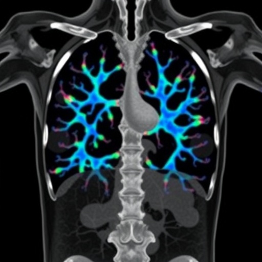
Enter radiomics—a sophisticated, data-driven approach employing advanced machine learning algorithms to quantitatively extract and analyze hundreds of imaging features invisible to the human eye. These features encapsulate complex textural, shape, and intensity attributes of lung tissue, providing a multidimensional characterization of pathological changes. The research team applied this technique to a trove of high-resolution CT scans from 320 sarcoidosis patients enrolled in the Genomic Research in Alpha-1 Antitrypsin Deficiency and Sarcoidosis (GRADS) Study, one of the most comprehensive sarcoidosis cohorts to date.
Using unsupervised machine learning clustering algorithms, the investigators discerned four distinct radiomic phenotypes within the sarcoidosis population, each representing a unique constellation of imaging features. These clusters ranged from patients exhibiting minimal and localized lung abnormalities to those manifesting widespread inflammation and fibrotic remodeling. Intriguingly, these radiomic subgroups correlated strongly with quantitative lung function metrics, including measures of airflow obstruction and gas exchange impairment, independent of conventional radiological staging. This suggests that radiomics captures dimensions of disease severity and heterogeneity that standard imaging assessments overlook.
Dr. Tasha Fingerlin, vice chair of Immunology and Genomic Medicine at National Jewish Health and co-senior author of the paper, emphasized the transformative potential of this technology. She noted that radiomic profiling enables an unprecedented granular view of sarcoidosis pathology, transcending the coarse categorical labels traditionally used in clinical practice. This refinement affords clinicians a more precise tool to stratify patients according to underlying disease biology rather than solely radiographic appearance.
Moreover, Dr. Lisa Maier, chief of Environmental and Occupational Health Sciences and head of the World Association of Sarcoidosis and Granulomatous Disease (WASOG) Sarcoidosis Center of Excellence at National Jewish Health, highlighted that radiomic analysis offers scalability through automation. Because these computational workflows utilize open-source software and require minimal manual intervention, they promise rapid, reproducible assessments of large imaging datasets, a crucial advantage for busy clinical settings and multi-center trials. This automation holds particular promise for resource-limited regions where sarcoidosis imaging expertise is scarce.
The ability of radiomics to objectively quantify pathological features also opens doors for dynamic disease monitoring. By applying this method longitudinally, clinicians could track progression or regression of lung lesions with unprecedented sensitivity, tailoring treatment regimens in near real-time. This personalized medicine approach could mitigate risks associated with overtreatment or treatment delay, two persistent challenges in sarcoidosis management.
Another compelling aspect of this work is its potential role in clinical research. Sarcoidosis is notoriously heterogeneous, complicating patient selection and outcome measures in clinical trials. Radiomic biomarkers could serve as standardized imaging endpoints or help identify homogeneous patient subgroups, thereby enhancing trial design and interpretability. This could expedite the development of novel therapeutics targeting specific disease phenotypes.
Despite its promise, the authors acknowledge that integrating radiomics into routine care necessitates further validation. Larger, prospective studies are required to confirm these findings across diverse populations and imaging platforms. Additionally, harmonizing radiomic feature extraction methods and establishing consensus on clinically relevant imaging signatures will be critical to ensure reproducibility and regulatory acceptance.
Importantly, National Jewish Health’s longstanding recognition as a WASOG Center of Excellence underscores its leading role in sarcoidosis research and patient care. The center’s robust infrastructure and expertise uniquely position it to pioneer the translation of radiomic innovations from bench to bedside, offering hope of improved outcomes for this challenging disease.
This study exemplifies the expanding frontier where medical imaging converges with computational science, marking a paradigm shift toward precision pulmonology. By unveiling the hidden narrative embedded within lung scans, radiomics stands to decode the complexity of sarcoidosis, offering clearer diagnostic clarity, improved prognostication, and ultimately, more personalized therapies for patients worldwide.
As the medical community awaits broader clinical adoption, this research heralds a new era in which artificial intelligence augments human expertise—turning static images into dynamic reservoirs of diagnostic insight and reshaping the future of inflammatory lung disease management.
Subject of Research: Radiomic analysis and imaging phenotyping in sarcoidosis.
Article Title: Radiomic profiling of chest CT in a cohort of sarcoidosis cases.
News Publication Date: 18-Feb-2026.
Web References:
- Article: https://pubmed.ncbi.nlm.nih.gov/41708686/
- DOI: http://dx.doi.org/10.1038/s41598-026-39384-9
Keywords: Sarcoidosis, Radiomics, Chest CT, Lung Imaging, Machine Learning, Pulmonary Disease, Inflammatory Lung Disease, Fibrosis, Medical Imaging, Personalized Medicine, Computational Radiology, Lung Function.
