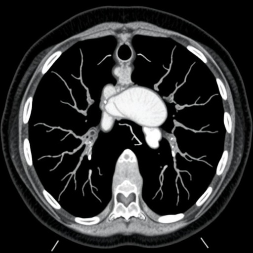
In an illuminating study published in Pediatric Radiology, a team of researchers led by Han et al. delves into the intricate realm of extralobar pulmonary sequestration (EPS) in children. This retrospective analysis highlights the computed tomography (CT) findings among a cohort of 58 pediatric patients, aiming to provide critical insights into this rare congenital lung condition. The researchers meticulously reviewed patient data, focusing on visualization techniques that illustrate the nuances of EPS.
Extralobar pulmonary sequestration is characterized by a non-functional lung tissue that is not connected to the bronchial tree and is often encased in its own pleura. Unlike the more common intralobar form, EPS is typically found outside the normal lung tissue, often supplied by aberrant systemic arteries. These unique vascular anatomies can lead to complex challenges in diagnosis and management, making advanced imaging techniques essential for precise assessment.
CT imaging plays a pivotal role in identifying extralobar sequestration. In their study, Han and colleagues meticulously categorized various CT findings, which included the location, size, and vascular supply of the sequestrated lung tissue. Notably, their analysis highlighted the importance of differentiating EPS from other similar lung anomalies. The distinction is critical because the management and surgical approaches may vary significantly between different types of pulmonary sequestrations.
The research team found that the most common presentation of EPS involved the lower lobes, although cases in the upper lobes were also documented. This distribution is essential for clinicians to understand, as it can influence surgical strategies and potential complications during interventions. Moreover, the relationship between the sequestrated tissue and surrounding lung structures illuminated the complexities involved in surgical planning, as well as the need for individualized patient evaluation.
An intriguing finding of the study was the frequent presence of associated anomalies in patients with EPS. These included congenital heart defects and other thoracic abnormalities. Such associations underscore the necessity for comprehensive imaging assessments, allowing for a greater understanding of a child’s overall health status and any potential impacts on treatment options. The researchers emphasized the need for a multidisciplinary approach during diagnosis and treatment to optimize patient outcomes.
The study also detailed the potential complications arising from inadequately addressed extralobar sequestration. Clinical manifestations can include recurrent infections, hemoptysis, and pneumothorax. Understanding these risks is vital for both pediatricians and radiologists, as timely intervention can significantly alter a child’s developmental trajectory and quality of life. The researchers noted that early identification through advanced imaging can provide significant advantages in the clinical management of these patients.
CT findings in cases of EPS often reveal diagnostic indicators that can cue radiologists into the condition. For example, some sequestrations displayed characteristic systemic arterial supply, which was highlighted with the use of contrast-enhanced imaging techniques. These findings not only assist in confirming the diagnosis but can also aid in preoperative planning. The authors urged for heightened awareness among healthcare professionals regarding these imaging features to facilitate earlier diagnosis and intervention.
Surgical resection remains the primary treatment approach for symptomatic extralobar pulmonary sequestration. The authors discussed various surgical techniques employed, from lobectomies to wedge resections, which vary based on the individualized anatomical and pathological considerations identified through imaging studies. Their exploration of surgical outcomes sheds light on the benefits of timely intervention and comprehensive pre-surgical planning—an essential aspect in improving postoperative results.
Through their in-depth analysis, Han et al. advocate for the integration of advanced imaging techniques in routine pediatric evaluations, particularly in cases with unexplained respiratory symptoms. The nuances of EPS call for diligence in diagnostic imaging and clinical assessment.
This study is a significant contribution to pediatric radiology literature, shedding light on the intricate interplay between congenital lung anomalies and imaging findings. The authors hope their findings will foster further research into EPS and underline the vital role of radiologists in the identification and management of this condition.
In conclusion, the retrospective study by Han and colleagues presents a compelling view of extralobar pulmonary sequestration, offering valuable insights that could lead to improved patient care. With detailed CT findings and a comprehensive understanding of associated anomalies, healthcare professionals are better equipped to handle this rare condition effectively.
While the road to recognizing and treating extralobar pulmonary sequestration remains complex, studies like this pave the way for optimized outcomes through better diagnostic practices and informed clinical decisions.
Subject of Research: Extralobar pulmonary sequestration in children
Article Title: Computed tomography findings of extralobar pulmonary sequestration in children: a retrospective study of 58 patients
Article References:
Han, Z., Yu, T., Duan, X. et al. Computed tomography findings of extralobar pulmonary sequestration in children: a retrospective study of 58 patients.
Pediatr Radiol 55, 1652–1668 (2025). https://doi.org/10.1007/s00247-025-06290-7
Image Credits: AI Generated
DOI:
Keywords: Extralobar pulmonary sequestration, pediatric radiology, computed tomography, congenital lung anomalies, surgical management, imaging findings.


