In a groundbreaking new study exploring the intricate relationships between metabolic health, brain structure, and cognitive function, researchers have uncovered that the cortical surface area (CSA) serves as a more potent intermediary than traditional metrics like body mass index (BMI) and waist-to-hip ratio (WHR) in connecting obesity-related measurements to cognitive impairment in patients experiencing their first episode of schizophrenia (FEPS). This discovery, published in BMC Psychiatry, shines a light on more nuanced biological underpinnings of cognitive deficits in schizophrenia and opens pathways for precision medicine strategies targeting metabolic and neuroanatomical factors.
Schizophrenia is a complex neuropsychiatric disorder characterized by profound cognitive impairments alongside psychotic symptoms. It is well established that metabolic disturbances frequently co-occur with schizophrenia, often exacerbating cognitive decline. However, until now, the mechanistic links between metabolic risk factors, brain structural changes, and cognitive performance remained elusive. The international research team, led by Lin et al., sought to dissect these associations by focusing on two key neuroanatomical metrics derived from advanced high-resolution 3.0-Tesla magnetic resonance imaging (MRI): cortical surface area and cortical thickness.
Involving a substantial cohort of 160 first-episode schizophrenia patients, alongside 150 healthy controls for comparison, the study incorporated a comprehensive evaluation of cognitive function using the Measurement and Treatment Research to Improve Cognition in Schizophrenia Consensus Cognitive Battery (MCCB). The psychiatric symptomatology was concurrently assessed by the Positive and Negative Syndrome Scale (PANSS). To capture the metabolic dimension, three obesity indices were scrutinized: BMI, waist-to-hip ratio, and waist-to-height ratio (WHtR), each reflecting different aspects of body composition and fat distribution.
One of the most striking revelations was that healthy controls exhibited significantly greater cortical surface area and thickness in multiple gray matter regions compared to their schizophrenia counterparts, even when accounting for confounding variables. This finding underscores a fundamental neuroanatomical deficit in the early stages of schizophrenia potentially linked to cognitive impairment. Crucially, among the metabolic parameters, WHtR emerged as a notably stronger correlate of cognitive performance relative to BMI and WHR, particularly in domains related to social cognition and overall MCCB composite scores.
Delving deeper, the analysis illuminated that bilateral cortical surface area acts as a mediator between both WHR and WHtR and cognitive outcomes. This mediating role implies that changes in the brain’s cortical morphology may bridge the detrimental effects of abdominal obesity and cognitive dysfunction. Among numerous brain regions analyzed, the right inferior parietal gyrus stood out as a critical hub where cortical surface area specifically mediated the relationship between obesity metrics and cognitive performance. This area is known for its role in integrating sensory information and contributing to higher-order cognitive processes, making its involvement especially relevant.
The study’s emphasis on waist-to-height ratio rather than more conventional obesity markers could signal a paradigm shift in how clinicians and researchers evaluate metabolic risks in schizophrenia. Unlike BMI, which does not differentiate muscle from fat mass or account for fat distribution, WHtR is a more precise gauge of central adiposity—an established risk factor for systemic inflammation and vascular pathology. Such factors may adversely affect brain health and cognition via pathways that compromise cerebrovascular integrity or promote neuroinflammation.
By honing in on cortical surface area, this research presents a compelling argument that morphological brain alterations may underpin the cognitive deficits linked with obesity in schizophrenia. Cortical thickness and surface area represent distinct neurodevelopmental properties: while thinning often reflects neurodegeneration, reductions in surface area might signal disrupted neurodevelopmental processes such as impaired synaptogenesis or dendritic arborization. The differential mediation effects observed point toward cortical surface area as an early and sensitive marker for the neurocognitive impact of metabolic disturbances.
Beyond its clinical implications, the study exemplifies the power of integrating neuroimaging with metabolic and cognitive assessments to unravel complex biopsychosocial interactions in psychiatric disorders. It suggests that targeting abdominal obesity could mitigate cognitive decline, potentially via neuroprotective interventions that preserve or restore cortical architecture. Furthermore, the right inferior parietal gyrus could represent a promising focal point for neuromodulatory treatments or cognitive rehabilitation programs aimed at enhancing function in vulnerable patient populations.
However, the authors also caution that their cross-sectional design limits causal interpretations, and future longitudinal studies are necessary to confirm the temporal sequence and potential reversibility of these alterations. Additional research should also probe molecular and cellular mechanisms linking adiposity, cortical morphometry, and cognition, including roles for inflammatory cytokines, insulin resistance, and neurotrophic factors.
This ambitious piece of research not only advances our understanding of schizophrenia’s multifaceted pathology but also underscores the importance of personalized medicine approaches that encompass somatic health to optimize psychiatric outcomes. Detecting and monitoring waist-to-height ratio could become a practical and non-invasive tool in routine psychiatric care, enabling earlier identification of patients at heightened risk for cognitive impairment and tailoring interventions accordingly.
As the global burden of metabolic syndrome and schizophrenia both continue to rise, dissecting the interplay between these conditions is critical. Lin et al.’s work offers a refined conceptual framework and methodological blueprint for future investigations aiming to unravel how peripheral health disturbances reverberate through brain structure to influence cognition and functioning.
In summary, this study highlights the superior role of cortical surface area, especially within the right inferior parietal gyrus, as a neuroanatomical mediator linking abdominal obesity—best captured by the waist-to-height ratio—to cognitive impairment in first-episode schizophrenia patients. It challenges prevailing reliance on BMI and waist-to-hip ratio, paving the way for enhanced biomarker development and integrative treatment strategies that address both brain and body health in severe mental illness.
Subject of Research: The study focuses on examining the mediating role of cortical surface area and cortical thickness in the association between obesity metrics (waist-to-height ratio, waist-to-hip ratio, body mass index) and cognitive impairment in patients with first-episode schizophrenia.
Article Title: Cortical surface area as a stronger mediator of the waist-to-height ratio and cognitive impairment link in patients with first-episode schizophrenia compared to body mass index and waist-hip ratio.
Article References: Lin, C., Yin, Y., Gou, M. et al. Cortical surface area as a stronger mediator of the waist-to-height ratio and cognitive impairment link in patients with first-episode schizophrenia compared to body mass index and waist-hip ratio. BMC Psychiatry 25, 962 (2025). https://doi.org/10.1186/s12888-025-07458-z
DOI: https://doi.org/10.1186/s12888-025-07458-z
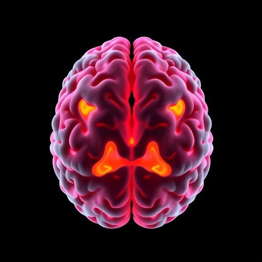
Image Credits: AI Generated
