


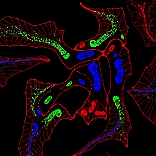
A molecular pathway long associated with immune suppression has emerged as a potential therapeutic target for capillary malformations, the congenital blood-vessel abnormalities that often appear as port-wine stains. In a study published in Angiogenesis, researchers at Amsterdam University Medical Centers and collaborating institutions traced how a mutation in the endothelial-cell signaling protein Gαq disrupts the behavior of developing blood vessels. Their experiments identified the Calcineurin–NFAT–DSCR1.4 signaling axis as a key point of failure, and showed that manipulating this circuit can restore important vascular-cell functions in laboratory models. The findings raise the possibility that medicines already used for other conditions could eventually be repurposed—or redesigned—to treat disorders such as isolated capillary malformations and Sturge-Weber syndrome. The work remains preclinical, but it offers one of the clearest mechanistic links yet between a disease-causing mutation and the abnormal movement and growth of endothelial cells.
Capillary malformations affect roughly three in every 1,000 newborns and are typically visible as persistent red or purple patches caused by enlarged, tortuous capillaries and post-capillary venules. For many people, the lesions are primarily a cosmetic concern, although they can thicken or darken over time and may cause tissue overgrowth. In some patients, particularly those with facial port-wine stains, the same underlying process is associated with Sturge-Weber syndrome. This neurocutaneous disorder can involve abnormal vessels in the eye and the membranes covering the brain, contributing to glaucoma, seizures, migraines and other neurological complications. Existing interventions—including pulsed-dye laser treatment, surgery and medicines to control eye pressure—can reduce symptoms or improve appearance, but they do not eliminate the abnormal endothelial cells that create the malformation. A treatment directed at the cellular machinery driving the lesions could therefore address a fundamental biological cause rather than only managing its consequences.
The genetic trigger in most cases is a somatic mutation in GNAQ, meaning that it arises after conception and is present only in a subset of cells rather than throughout the body. The most common alteration changes the amino acid arginine to glutamine at position 183 of the Gαq protein, a substitution known as p.R183Q. Gαq normally functions as part of a molecular relay downstream of G protein-coupled receptors. When activated, it stimulates phospholipase C-β, an enzyme that cleaves the membrane lipid PIP2 into two signaling molecules: diacylglycerol and inositol 1,4,5-trisphosphate, or IP3. IP3 releases calcium from intracellular stores, while diacylglycerol activates protein kinase C. Together, these signals influence cell growth, survival, shape and migration. The R183Q mutant appears to keep this relay abnormally active, producing persistent calcium signaling and confusing the instructions that endothelial cells use to organize blood vessels.
To isolate the mutation’s effects, the researchers used CRISPR–Cas9 gene editing to remove endogenous Gαq from immortalized human dermal microvascular endothelial cells. They then reintroduced either normal Gαq or the R183Q mutant using lentiviral vectors. This design allowed the two cell populations to be compared while keeping much of their genetic and environmental background the same. The engineered proteins carried a fluorescent mTurquoise2 tag, enabling the investigators to confirm their expression and localization. Although both rescued cell lines produced similar amounts of Gαq—at levels approximately three times higher than those normally found in the parental cells—the mutant cells developed a distinctly altered shape. They became larger and more elongated, even though their VE-cadherin cell junctions and actin cytoskeleton remained broadly organized. The change in morphology was accompanied by a functional defect: in scratch-wound experiments, mutant endothelial cells moved slowly into the cleared area, leaving the artificial wound open for longer than cells carrying normal Gαq.
The researchers also tested angiogenic sprouting, a process in which endothelial cells extend coordinated projections to form new vessel-like structures. They compacted the cells into three-dimensional spheroids, embedded them in collagen and stimulated them with vascular endothelial growth factor, or VEGF. Compared with normal-Gαq spheroids, those expressing Gαq-R183Q produced substantially fewer and shorter sprouts. This result is notable because vascular malformations are not simply caused by excessive vessel growth; they can also arise when endothelial cells fail to migrate, interpret developmental signals or remodel their surroundings correctly. The mutant cells’ poor performance in both migration and sprouting assays suggested that the mutation was disrupting a network of intracellular signals required for orderly vascular development rather than merely accelerating proliferation. The two cell populations showed no major difference in proliferation rates, further focusing attention on signaling and cell behavior.
To map those signaling changes without presupposing which pathway was involved, the team turned to quantitative phosphoproteomics. Proteins are often regulated by phosphorylation, the reversible addition of phosphate groups to specific amino acids. By measuring thousands of phosphorylation sites, scientists can build a broad picture of which molecular circuits are active or suppressed inside a cell. The researchers grew the normal and mutant endothelial cells in media containing either ordinary amino acids or isotopically heavy versions of lysine and arginine, a strategy called SILAC. After mixing cell lysates, they used mass spectrometry to distinguish the two populations and enriched phosphorylated peptides for analysis. Label-swap experiments helped control for technical bias. Pathway analysis highlighted increased activity associated with the phosphatase PTEN and reduced activity in the integrin-linked kinase pathway. Immunoblotting confirmed lower phosphorylation of Akt, S6K and Paxillin, molecules involved in survival, growth and adhesion, while phosphorylation of ERK was increased, consistent with earlier studies of the mutation.
The strongest signal, however, involved Calcineurin and the NFAT family of transcription factors. Calcineurin is a calcium- and calmodulin-dependent serine/threonine phosphatase. When intracellular calcium rises, Calcineurin removes phosphate groups from NFAT proteins, allowing them to move from the cytoplasm into the nucleus and regulate gene expression. In the mutant endothelial cells, several phosphorylation sites on NFAT1 and NFAT2 were reduced, indicating enhanced dephosphorylation. Yet imaging revealed an apparent paradox: rather than accumulating in the nucleus, both NFAT proteins remained predominantly in the cytoplasm. The explanation appeared to be a feedback inhibitor called DSCR1.4, also known as RCAN1.4. Its abundance rose sharply in the Gαq-R183Q cells, whereas the related DSCR1.1 isoform did not change. DSCR1.4 can inhibit Calcineurin, creating a feedback loop in which excessive upstream signaling ultimately suppresses the nuclear NFAT activity needed for normal endothelial responses. The mutant cells therefore showed biochemical evidence of persistent pathway stimulation alongside a failure of NFAT-dependent signaling in the nucleus.
Patient tissue provided an important reality check. In skin biopsies from people whose capillary malformations carried the GNAQ p.R183Q mutation, immunofluorescence microscopy showed NFAT1 and NFAT2 concentrated mainly in the cytoplasm of VE-cadherin-positive endothelial cells. Phosphorylated NFAT signals that were detectable in experimental controls were not observed in the endothelial cells of the malformation samples, and DSCR1 protein was present in the affected tissue. These observations were consistent with the engineered-cell experiments, although the researchers noted a significant limitation: matched healthy skin biopsies were not available for direct comparison. The patient samples therefore support the proposed signaling defect but cannot by themselves establish how much NFAT localization or DSCR1.4 expression differs from normal capillaries. Even so, the alignment between patient tissue and the laboratory model strengthens the case that the pathway is relevant to the disease rather than being an artifact of cell culture.
The team next asked whether the pathway could be pharmacologically manipulated. They treated mutant endothelial cells with tacrolimus, also called FK506, an immunosuppressive drug that inhibits Calcineurin after binding to FKBP proteins. A 24-hour treatment restored the nuclear-to-cytoplasmic distribution of NFAT1 and NFAT2 toward the pattern seen in normal cells. It also restored phosphorylation at the NFAT1 and NFAT2 sites that had been depleted by the mutation. Functionally, tacrolimus slightly improved wound closure and partially rescued the number and length of VEGF-induced sprouts. The effect was real but incomplete. The investigators suggest that mutant cells express lower levels of several FKBP proteins needed for tacrolimus to inhibit Calcineurin efficiently, and that additional signaling pathways remain active downstream of Gαq-R183Q. The result is therefore not evidence that tacrolimus is ready to treat capillary malformations, particularly because systemic immunosuppression carries substantial risks, but it does demonstrate that the abnormal pathway is chemically accessible.
A more dramatic rescue came from targeting DSCR1 directly. Using a lentiviral short-hairpin RNA, the researchers reduced expression of both DSCR1.1 and DSCR1.4 in the engineered cells. In Gαq-R183Q endothelial cells, DSCR1 depletion increased phosphorylation of NFAT1 and NFAT2, promoted their movement into the nucleus and almost completely restored migration in scratch assays. It also strongly enhanced angiogenic sprouting in the three-dimensional collagen model. The contrast with tacrolimus is mechanistically informative: blocking Calcineurin from outside the cell produced only a partial functional recovery, whereas removing the mutation-induced feedback brake had a much larger effect. The result identifies DSCR1.4 as a particularly attractive target, although suppressing a gene throughout the body would be technically and biologically challenging. Future therapies might need to deliver a DSCR1.4-directed treatment selectively to affected endothelial cells or develop molecules that interrupt its interaction with Calcineurin without disturbing essential signaling elsewhere.
The findings place capillary malformations within a growing landscape of genetically defined vascular disorders in which different mutations converge on distinct but interconnected signaling networks. The study found reduced Akt/mTOR activity in Gαq-R183Q cells, in contrast to other malformations driven by activating mutations in PIK3CA or loss of PTEN function, where the same pathway is often overactive. That difference underscores why treatments cannot necessarily be transferred from one vascular anomaly to another simply because the lesions look similar. The researchers also caution that their model used cultured human endothelial cells expressing the mutant protein at higher-than-endogenous levels, and that the experiments did not reproduce the full complexity of skin, brain or eye tissue. Animal studies and disease-relevant human models will be needed to determine whether selectively correcting Calcineurin–NFAT–DSCR1.4 signaling can shrink established lesions, prevent progression or improve neurological and ophthalmological complications. Still, the work offers a compelling new route from mutation to mechanism to therapeutic strategy—and suggests that the molecular circuitry beneath a visible birthmark may be far more precisely targetable than previously thought.
Cite Scienmag News
Nathaniel Bowman. (August 27, 2026). Calcineurin-NFAT-DSCR1.4 Emerges as Drug Target in Gαq-R183Q-Driven Capillary Malformations. Scienmag. https://scienmag.com/calcineurin-nfat-dscr1-4-emerges-as-drug-target-in-g%ce%b1q-r183q-driven-capillary-malformations/
Nathaniel Bowman. "Calcineurin-NFAT-DSCR1.4 Emerges as Drug Target in Gαq-R183Q-Driven Capillary Malformations." Scienmag, 27 August 2026, https://scienmag.com/calcineurin-nfat-dscr1-4-emerges-as-drug-target-in-g%ce%b1q-r183q-driven-capillary-malformations/. Accessed 6 September 2026.
Nathaniel Bowman. "Calcineurin-NFAT-DSCR1.4 Emerges as Drug Target in Gαq-R183Q-Driven Capillary Malformations." Scienmag. August 27, 2026. https://scienmag.com/calcineurin-nfat-dscr1-4-emerges-as-drug-target-in-g%ce%b1q-r183q-driven-capillary-malformations/


