In a groundbreaking study published recently in Nature Neuroscience, researchers have uncovered a previously uncharted neural pathway that links environmental light exposure to the regulation of feeding behavior and body weight. This finding has significant implications for understanding the subtle yet profound influence of light on metabolic functions and could pave the way for novel interventions targeting obesity and related disorders. By pinpointing a specific visual circuit involving retinal neurons and the lateral hypothalamus, the study elucidates the mechanistic foundation behind the long-observed effects of bright light therapy on appetite suppression and weight management.
The interplay between circadian rhythms, environmental light, and metabolic processes has long intrigued scientists. While the influence of light on sleep-wake cycles is well-established, nonimage-forming aspects of light perception—such as its impact on feeding behavior—remain less clearly defined. Prior evidence suggested that exposure to bright light could curb appetite and reduce weight gain, yet the precise neuronal substrates responsible for these effects were obscure. The research team, led by Li, Huang, and Xu, sought to bridge this knowledge gap by investigating the role of specific retinal ganglion cells and hypothalamic circuits in mediating the metabolic responses to light.
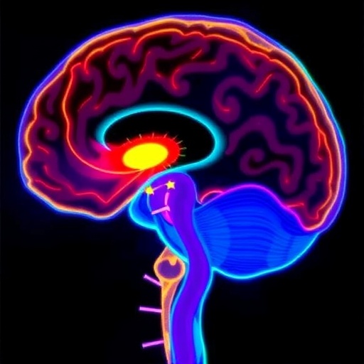
At the core of this discovery is a subset of ON-type retinal ganglion cells that express the neurofilament marker SMI-32. These retinal ganglion cells act as sensitive detectors of bright light and send visual signals directly to the ventral lateral geniculate nucleus (vLGN), a thalamic region not traditionally associated with feeding regulation. The vLGN, enriched with GABAergic neurons, functions as a relay hub. These neurons exert inhibitory control over GABAergic neurons residing in the lateral hypothalamic area (LHA), a brain region historically implicated in energy balance, feeding, and reward processing.
The lateral hypothalamus has long been recognized as a key player in orchestrating feeding responses, but the new study reveals that its activity can be modulated by superior visual inputs beyond the classical image-forming pathways. Researchers demonstrated that bright light stimulates the retinal ganglion cells projecting to the vLGN, which in turn suppresses LHA GABAergic neurons. This suppression decreases downstream signaling that promotes food intake, effectively curbing appetite. This neural cascade delineates an elegant feedback loop through which environmental light can exert a powerful metabolic influence.
Crucially, the investigators employed sophisticated techniques such as optogenetics to specifically activate both the vLGN-projecting retinal ganglion cells and the vLGN-to-LHA neuronal projections. Activation of these pathways alone was sufficient to induce a marked reduction in food consumption and to attenuate weight gain in murine models. This approach enabled the team to dissect the causative chain from retinal stimulation all the way to hypothalamic control of feeding, thus providing direct evidence of the necessity and sufficiency of this visual circuit for the feeding-suppressive effects of bright light exposure.
Intriguingly, the findings reconcile earlier clinical observations of bright light therapy’s beneficial role in weight management, often seen in human studies addressing seasonal affective disorder and obesity. By pinpointing the retina–vLGN–LHA axis, the study offers a neurobiological substrate that could underpin these therapeutic effects. It invites deeper investigation into how circadian lighting environments might be optimized to leverage this natural appetite-suppressing circuitry.
The study also highlights a critical role of GABAergic neurons in both the vLGN and LHA, suggesting a complex inhibitory network that fine-tunes feeding behavior in response to photic signals. The GABA neurotransmitter, known for its inhibitory functions, acts within this circuit to modulate neuronal excitability and behavior. This interplay provides a new dimension to understanding how sensory input can translate into metabolic outcomes through discrete, defined inhibitory pathways.
Furthermore, the research underscores the specificity of retinal ganglion cell subtypes in mediating non-image forming physiological responses. While ipRGCs (intrinsically photosensitive retinal ganglion cells) have been implicated in circadian regulation, this study draws attention to SMI-32-expressing ON-type retinal ganglion cells as pivotal in feeding-related outcomes, expanding the functional diversity of retinal pathways beyond classical roles.
This discovery opens doors to several translational prospects. For instance, designing light-based therapies or devices that target this visual circuit could complement pharmacological approaches to obesity. Additionally, understanding the diurnal variations in this circuit could inform behavioral strategies for weight control, leveraging timed light exposure to maximize metabolic benefits.
Importantly, the study prompts a reconsideration of the lateral hypothalamic area’s function beyond its traditional behavioral roles. It exemplifies how sensory input integration, particularly from environmental light, can dynamically modulate hypothalamic circuits governing energy homeostasis, revealing a sophisticated neural network that bridges external stimuli and internal physiological states.
Overall, the identification of the retina–vLGN–LHA circuit expands our understanding of the visual system’s influence on feeding beyond image perception, positioning light as a potent modulator of metabolic health. This intersection of sensory neuroscience and metabolic regulation is poised to inspire further research into environmental factors shaping human health in profound, previously unappreciated ways.
As obesity rates worldwide continue to rise, innovative strategies addressing its complex neurobiological underpinnings are urgently needed. This study’s insights into a defined visual circuit that suppresses feeding and weight gain via bright light exposure provide a compelling avenue for non-invasive interventions. It also encourages a broader perspective on how daily environmental factors, such as light, intricately sculpt behavior and physiology.
Future investigations may explore how this circuit interacts with other hypothalamic pathways controlling hunger, satiety, and energy expenditure. Additionally, it will be critical to determine whether similar mechanisms operate in humans and how individual differences in retinal or hypothalamic function might influence responsiveness to bright light-based therapies.
In summary, the research by Li, Huang, Xu, and colleagues represents a milestone in decoding the neural pathways linking environmental cues to fundamental survival behaviors like feeding. It highlights the capacity of the nervous system to leverage sensory information, converting light signals into neurochemical instructions that shape energy balance—a testament to the complexity and elegance of brain function.
The visual circuit delineated in this study not only answers longstanding questions about light’s metabolic influence but also reframes therapeutic possibilities, urging a multidisciplinary approach intertwining neuroscience, endocrinology, and environmental health sciences. This confluence promises to enrich our arsenal against obesity while deepening appreciation for the brain’s integrative role in adapting to the environment.
Subject of Research: Mechanisms by which bright light exposure suppresses feeding and weight gain through a visual neural circuit involving the retina, ventral lateral geniculate nucleus, and lateral hypothalamic area.
Article Title: Bright light exposure suppresses feeding and weight gain via a visual circuit linked to the lateral hypothalamus.
Article References:
Li, W., Huang, X., Xu, X. et al. Bright light exposure suppresses feeding and weight gain via a visual circuit linked to the lateral hypothalamus. Nat Neurosci (2025). https://doi.org/10.1038/s41593-025-02156-1
Image Credits: AI Generated
