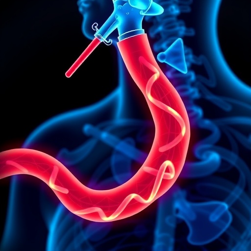
A groundbreaking advancement in the early detection of esophageal cancer has emerged through the integration of two cutting-edge imaging modalities into a single, innovative capsule endoscopy device. Esophageal cancer remains one of the deadliest malignancies worldwide, largely due to its typically late diagnosis, which drastically diminishes survival rates. While late-stage esophageal cancer carries a survival rate near ten percent, early diagnosis improves patient prognosis dramatically, raising survival chances to around ninety percent. This stark contrast underscores the critical need for technologies capable of identifying subtle, pre-cancerous tissue alterations before the disease progresses. The new O2E technology, developed by a collaborative team of biomedical engineers and clinicians, represents a significant leap toward this goal by enabling unprecedented visualization of the esophageal mucosa and submucosa at microscopic levels.
The core innovation of the O2E capsule rests in its dual-imaging system, which synergistically combines optical coherence tomography (OCT) with optoacoustic (photoacoustic) imaging. OCT is a well-established modality famed for its ability to generate high-resolution cross-sectional images of tissue architecture by measuring the backscattering of near-infrared light. However, OCT traditionally lacks sensitivity to vascular features beneath the surface layers. To augment this, the O2E system incorporates optoacoustic imaging, a technique that employs short laser pulses to induce thermoelastic expansion in blood vessels, generating ultrasound waves that can be detected externally. This modality is extremely sensitive to hemoglobin absorption, enabling detailed visualization of microvascular networks several millimeters beneath the tissue surface — a vital indicator of neoplastic transformation.
What makes this technology revolutionary is how both imaging modalities are seamlessly integrated into a miniaturized, tethered capsule capable of scanning the esophagus in a full 360-degree field of view. The capsule is designed to be navigated through the esophagus, capturing volumetric data sets that reveal not only the microstructural organization of the tissue but also the functional state of the vasculature in exquisitely high spatial resolution. This comprehensive imaging capability allows clinicians to detect minuscule changes in tissue morphology and blood vessel formation associated with the earliest stages of esophageal neoplasia, changes that conventional endoscopy or imaging have failingly missed.
The significance of imaging microvascular features lies in the fact that angiogenesis – the formation of new blood vessels – is an early hallmark of malignant transformation in many cancers. Prior to this, subtle microvascular remodeling beneath the epithelial surface was difficult to assess without invasive biopsies or contrast agents. The label-free nature of optoacoustic imaging, combined with OCT’s structural insights, allows a holistic characterization of tissue pathology in real-time. As Prof. Vasilis Ntziachristos, a pioneer in biomedical imaging and director at Helmholtz Munich, states, this dual imaging strategy exposes hidden features of early cancerous lesions, providing a window into previously inaccessible biophysical changes within the esophageal lining.
To validate their innovative imaging concept, researchers conducted pilot studies involving animal esophageal tissues as well as human biopsy specimens from patients diagnosed with Barrett’s esophagus—a recognized precursor to esophageal adenocarcinoma. The findings were compelling: the system reliably differentiated between healthy mucosa, tissue exhibiting dysplasia, and fully developed malignancies. The juxtaposition of structural OCT images alongside optoacoustic vascular maps offered an unparalleled differentiation capability, potentially allowing clinicians to pinpoint areas warranting closer examination or targeted biopsy.
In a striking initial demonstration, the research team tested the O2E capsule in vivo by scanning the inner lip mucosa of healthy volunteers. The choice of lip tissue was strategic due to its histological similarities to the esophagus in terms of stratified squamous epithelium and vascular architecture. These early human trials confirmed the capsule’s ethical safety and functionality, laying the groundwork for subsequent studies directly targeting esophageal visualization.
Looking forward, the project funded under the auspices of the European Innovation Council (EIC) Pathfinder initiative, named ESOHISTO and launched in 2025, aims to refine this promising technology toward clinical application. Developing a system robust enough for routine use in clinical endoscopy suites involves tackling challenges such as miniaturization of components, real-time data processing, and ergonomic capsule designs compatible with patient comfort and clinical workflow. Increasingly sophisticated algorithms will also be explored to automate image interpretation to aid gastroenterologists’ diagnostic confidence.
One particularly exciting direction is the planned integration of confocal endomicroscopy within the capsule platform. Confocal endomicroscopy utilizes focused light to image living tissue at cellular resolutions in vivo, allowing real-time microscopic assessment of cellular morphology. When combined with OCT and optoacoustic imaging, confocal capabilities could transform diagnostics by enabling simultaneous visualization of tissue architecture, vascularity, and cellular details. This multimodal approach heralds a new era of high-resolution, label-free molecular endoscopy that could precisely pinpoint molecular markers indicative of malignancy and thus revolutionize personalized cancer management.
The anticipated clinical impact extends beyond diagnostics. Current esophageal cancer staging and treatment often necessitate multiple biopsies, which carry risks such as bleeding, infection, and sampling errors. The enhanced sensitivity and specificity of this capsule endoscopy system could considerably reduce the requirement for invasive biopsies, accelerating diagnosis and facilitating timely therapeutic interventions. Moreover, early-stage cancer treatment is markedly less expensive and more effective than managing advanced disease, offering a compelling argument for broad healthcare implementation.
Economic considerations underline the urgency of such technologies. Treating advanced esophageal cancer patients can incur costs upward of 140,000 euros per individual, encompassing surgery, chemotherapy, radiotherapy, and prolonged hospital stays. Conversely, early detection and intervention could reduce these expenditures to approximately 10,000 euros, representing profound savings for healthcare systems while simultaneously improving quality of life and survival for patients. The implementation of O2E technology, therefore, stands to contribute substantially to healthcare sustainability while dramatically transforming patient outcomes.
Helmholtz Munich, a leading institution driving this innovation, exemplifies the fusion of engineering and biomedical science. With around 2,500 employees and part of the broader Helmholtz Association – Germany’s largest scientific organization – the center focuses on interdisciplinary research that bridges bioengineering, artificial intelligence, and clinical medicine to tackle a spectrum of complex diseases. The collaborative nature of this research, involving specialists in imaging physics, molecular biology, and clinical oncology, has been crucial in realizing a technology with such translational potential.
Dr. Qian Li from the Medical University of Vienna, first author of the underlying study, emphasizes the transformative nature of this research on esophageal pathology diagnostics. The ambition is to evolve disruptions in imaging into practical tools that not only detect disease earlier but also inform therapeutic decision-making through high-resolution, molecularly targeted imaging. This represents a paradigm shift in endoscopic technology, moving it from purely visual assessment toward sophisticated molecular characterization.
The recent publication of their study in the reputed journal Nature Biomedical Engineering on August 6, 2025, marks an important milestone in the dissemination of this knowledge to the global scientific community. The work titled “Tethered optoacoustic and optical coherence tomography capsule endoscopy for label-free assessment of Barrett’s oesophageal neoplasia” stands as a beacon for future research and clinical translation. It is a clarion call for continued interdisciplinary collaboration to refine, validate, and deploy this powerful imaging platform widely.
In summary, the O2E capsule endoscopy system introduces a novel, label-free approach that merges structural and functional imaging to reveal hidden early-stage esophageal cancer biomarkers. Its ability to deliver comprehensive, high-resolution 3D images of both tissue microarchitecture and microvascular alterations offers a critical advantage over existing diagnostic tools. As the technology advances through refinement and clinical validation under the ESOHISTO project, it holds promise to radically transform esophageal cancer diagnostics, reduce patient morbidity, and substantially alleviate healthcare burdens worldwide. This convergence of engineering excellence and biomedical inquiry heralds a promising future in the fight against esophageal cancer.
Subject of Research: Early detection of esophageal neoplasia using dual-modality imaging capsule endoscopy combining optical coherence tomography and optoacoustic imaging.
Article Title: ‘Tethered optoacoustic and optical coherence tomography capsule endoscopy for label-free assessment of Barrett’s oesophageal neoplasia’
News Publication Date: 6-Aug-2025
Web References: DOI 10.1038/s41551-025-01462-0
Image Credits: Helmholtz Munich / Christian Zakian
Keywords: Esophageal cancer, Barrett’s esophagus, optical coherence tomography, optoacoustic imaging, capsule endoscopy, label-free imaging, early cancer detection, microvascular imaging, confocal endomicroscopy, translational biomedical imaging, biomedical engineering, minimally invasive diagnostics


