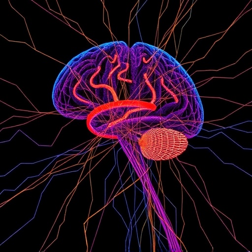
In a groundbreaking advancement poised to revolutionize neuroimaging, researchers have unveiled a cutting-edge method called Computational Scattered Light Imaging (ComSLI), setting a new benchmark for detailed mapping of nerve fiber networks within preserved brain tissues. This novel technique surmounts longstanding challenges in visualizing intricate neuronal pathways in brain slices embedded in paraffin wax—a standard preservation method—ushering in new possibilities for both neurological research and clinical diagnostics.
Understanding the complex architecture of the brain’s nerve fibers is fundamental to untangling the underpinnings of neurological disorders, including Alzheimer’s, Parkinson’s, and multiple sclerosis. Traditionally, brain tissues are immersed in paraffin wax to facilitate the creation of ultra-thin sections for microscopic examination, known as formalin-fixed paraffin-embedded (FFPE) sections. Despite the widespread use of FFPE samples in neuroscience and pathology, accurately charting the densely interwoven nerve fibers within these sections has been virtually impossible due to their optical properties and the limitations of conventional microscopy techniques.
The development of ComSLI represents a milestone achieved through international collaboration, involving physicists and neuroscientists from Delft University of Technology, Stanford University, Forschungszentrum Jülich, and Erasmus MC Rotterdam. Spearheaded by physicist Miriam Menzel, ComSLI harnesses the interaction of rotationally scattered LED light and computational imaging to reveal nerve fiber configurations with micrometer-scale precision, capturing both the breadth and detail of neuronal networks across substantial tissue areas.
ComSLI operates by illuminating a thin histological section from beneath with a rotating LED light source. This light permeates the tissue and is scattered by microscopic structures like nerve fibers. A high-resolution camera positioned above captures the scattered patterns, and sophisticated algorithms reconstruct these light interactions into detailed fiber maps. Unlike traditional microscopy that relies heavily on staining or fluorescence, ComSLI exploits intrinsic light scattering properties, enabling label-free, non-destructive visualization in a range of tissue preparations.
One of the most remarkable aspects of ComSLI is its versatility. The system functions with all common histological samples, including fresh-frozen and chemically fixed tissues, regardless of staining protocols or archival age. This feature means that priceless collections containing century-old brain slices can be re-examined retrospectively, injecting new life into existing tissue banks and enhancing our understanding of historical neuropathological cases.
The impact of ComSLI extends beyond methodological innovation. By applying ComSLI to the renowned BigBrain project—a comprehensive three-dimensional human brain atlas constructed from thousands of FFPE sections—the team demonstrated the technique’s power to parallel the well-delineated cellular architecture with its equally complex and previously elusive nerve fiber networks. This complementary visualization paves the way for integrated brain atlases that reveal not only cellular distributions but also the connectivity that orchestrates brain function.
From a practical standpoint, ComSLI’s hardware requirements are refreshingly modest: a rotating LED light source and a high-resolution camera. This simplicity significantly lowers barriers to adoption, enabling laboratories worldwide to implement the technique either as standalone systems or as cost-effective add-ons to existing microscopes. As a result, ComSLI could rapidly disseminate, democratizing high-precision nerve fiber mapping.
The clinical potential of ComSLI is equally promising. The ability to map disorganized nerve fibers within neurodegenerative tissue samples offers a new window into disease progression and pathology. Additionally, ComSLI’s proficiency in imaging fibrous structures beyond the nervous system, such as muscle and collagen fibers, extends its applicability into oncology. Surgeons could leverage fresh-frozen samples intra-operatively to assess tumor margins through collagen organization, enhancing surgical precision and outcomes.
ComSLI’s innovative approach leverages advances in computational imaging and light scattering physics, marking a convergence of interdisciplinary fields. Its capacity to accurately resolve fiber orientations and densities with micron resolution could catalyze breakthroughs in understanding how microstructural changes correlate with functional deficits in brain disorders.
This technology situates itself within the broader landscape of imaging physics, a domain where Delft University of Technology stands as a global leader. The university’s Imaging Physics department has a storied history of pioneering innovations that harness physical principles to develop transformative imaging modalities, impacting healthcare and digital society alike.
Looking ahead, ComSLI’s integration into neuropathology workflows could transform diagnostic paradigms. By providing label-free, high-resolution fiber maps, it may accelerate biomarker discovery and enable nuanced phenotyping of neurological diseases, ultimately guiding therapeutic interventions. Moreover, its compatibility with archived samples opens vast retrospective research avenues, potentially rewriting our understanding of disease mechanisms.
Summarily, Computational Scattered Light Imaging embodies a significant leap in neurohistological imaging, enabling comprehensive, precise mapping of nerve fibers in preserved human brain tissues. Its accessibility, versatility, and broad applicability position ComSLI as a powerful tool destined to invigorate both research and clinical spheres in neuroscience and beyond.
Subject of Research: Human tissue samples
Article Title: Micron-resolution fiber mapping in histology independent of sample preparation
News Publication Date: 5-Nov-2025
Web References:
DOI link to article
BigBrain atlas
Menzel Lab
Convergence Imaging Facility and Innovation Centre (CIFIC)
Image Credits: ScienceBrush
Keywords: Computational Scattered Light Imaging, ComSLI, nerve fiber mapping, FFPE brain sections, neuroimaging, microscopy, paraffin-embedded tissue, brain atlas, BigBrain, high-resolution imaging, neurological disorders, imaging physics


