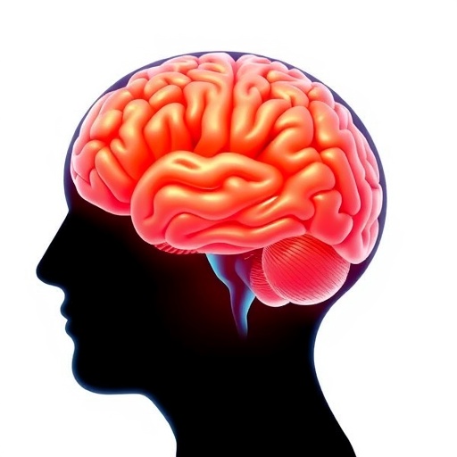
In a groundbreaking study poised to shift the paradigm of Parkinson’s disease (PD) research and treatment, scientists have unveiled critical insights into the structural and functional brain alterations that underpin pain perception in Parkinson’s patients. This latest research, published in the esteemed journal npj Parkinsons Disease, dissects the intricate neurological changes associated with pain, a symptom often overshadowed by the motor dysfunctions traditionally linked with PD. By exploring the multifaceted brain mechanisms involved, the study not only elevates our understanding of pain within this neurodegenerative disorder but also opens new avenues for targeted therapeutic interventions.
Parkinson’s disease has long been characterized predominantly by its motor symptoms—tremor, rigidity, bradykinesia—but non-motor symptoms like pain have increasingly gained attention for their profound impact on patient quality of life. Pain, a frequent yet enigmatic symptom in PD, is often underrecognized and undertreated. The study spearheaded by Wang, Jia, Yuan, and colleagues takes a deep dive into both structural and functional neuroimaging modalities to explore how changes in brain anatomy and network connectivity may contribute to pain sensations experienced by individuals with Parkinson’s.
Using advanced neuroimaging techniques including structural MRI and functional MRI, the research team conducted a comprehensive analysis involving a cohort of PD patients suffering from chronic pain alongside matched controls. Their rigorous methodology allowed for the simultaneous examination of gray matter volume, white matter integrity, and dynamic brain activity patterns during rest and pain-inducing stimuli. This dual approach was instrumental in teasing apart the disparate yet interrelated neural circuits that integrate sensory, emotional, and cognitive dimensions of pain.
One of the study’s pivotal findings is the identification of significant atrophy in brain regions traditionally implicated in pain processing, such as the insular cortex, anterior cingulate cortex, and thalamus. These areas exhibited reduced gray matter density correlated strongly with subjective pain ratings. This neurodegeneration appears to disrupt normal pain modulation pathways, possibly exacerbating the chronic pain states often reported by Parkinson’s patients. These results underscore the role of neuroanatomical deterioration beyond the classical dopaminergic deficits seen in motor symptom development.
Complementing these structural insights, the functional MRI data unveiled aberrations in resting-state connectivity within the brain’s pain matrix. In particular, disrupted communication was noted between the prefrontal cortex and limbic structures, regions critical for the integration of cognitive and emotional aspects of pain. Such functional disconnections might help explain why pain in PD is frequently accompanied by heightened emotional distress and cognitive interference, contributing substantially to patient suffering and disability.
Importantly, the study also highlighted alterations in white matter tracts that connect key hubs in the pain processing network. Diffusion tensor imaging metrics revealed reduced fractional anisotropy in several pathways including the spinothalamic tract and fronto-limbic circuits, indicating microstructural damage. These findings suggest that disrupted pathways at multiple levels—from peripheral sensory relay to higher-order integrative centers—collectively contribute to the complex pain phenotype in Parkinson’s disease.
Beyond mapping brain abnormalities, the investigators probed the neurochemical milieu using positron emission tomography (PET) imaging to quantify changes in neurotransmitter systems intricately tied to pain modulation. Notably, they observed diminished dopaminergic activity in areas implicated in both motor control and pain perception, reinforcing the dual role of dopamine in these processes. Additionally, alterations in serotonergic and opioidergic signaling were detected, which may reflect compensatory or maladaptive neural responses influencing pain thresholds.
This multipronged approach allowed the researchers to construct a comprehensive neurobiological profile of pain in Parkinson’s disease, situating it within a broader network of structural degeneration, functional dysconnectivity, and neurotransmitter imbalance. The convergence of these pathological factors likely accounts for the heterogeneity and persistence of pain symptoms among patients, emphasizing why conventional analgesics often fail to provide adequate relief.
The implications of these findings extend far beyond the academic sphere, holding promise for clinical translation. By delineating specific brain targets involved in PD-related pain, this study paves the way for more precise diagnostic tools and personalized treatment modalities. Neuromodulation techniques such as transcranial magnetic stimulation or deep brain stimulation might be refined to target these newly identified circuits, while pharmacological strategies could evolve to address the neurochemical disturbances unique to Parkinsonian pain.
Moreover, recognizing pain as a core feature of Parkinson’s disease necessitates a paradigm shift in clinical management, wherein neurologists assess and treat pain proactively alongside motor symptoms. The neuroimaging biomarkers uncovered here could aid in stratifying patients according to their pain risk and tailoring interventions accordingly, enhancing overall therapeutic outcomes.
The study also invites further research to unravel the dynamic interplay between neurodegeneration, neuroinflammation, and functional plasticity in shaping pain experiences. Longitudinal investigations tracking these brain alterations over the disease course would be invaluable in understanding the progression of pain and identifying critical windows for intervention.
Beyond Parkinson’s, the methodological framework applied in this research serves as a model for exploring pain in other neurodegenerative disorders, highlighting shared and distinct mechanisms across diseases. The integration of multimodal imaging with clinical phenotyping exemplifies how modern neuroscience can elucidate complex symptomatology that transcends traditional disease boundaries.
In sum, the work of Wang and colleagues represents a landmark contribution by illuminating the neurobiological underpinnings of pain in Parkinson’s disease with unprecedented detail and sophistication. It not only enriches our scientific comprehension but also lays foundational knowledge for developing holistic and effective care strategies. Patients with Parkinson’s, who often endure pain silently, can hope for a future where their suffering is recognized, understood, and effectively treated thanks to such innovative research endeavors.
As the neuroscience community builds upon these findings, the prospect of transforming pain management in Parkinson’s disease from an art of trial and error into a science of precision comes closer to reality. This study marks a critical step toward unraveling the enigmatic curse of pain in Parkinson’s and ultimately improving the lives of millions affected worldwide.
Subject of Research: Structural and functional brain alterations associated with pain in Parkinson’s disease
Article Title: Structural and functional brain alterations associated with pain in Parkinson’s disease
Article References:
Wang, E., Jia, Y., Yuan, P. et al. Structural and functional brain alterations associated with pain in Parkinson’s disease. npj Parkinsons Dis. (2025). https://doi.org/10.1038/s41531-025-01210-w
Image Credits: AI Generated
