In a groundbreaking study recently published in BMC Psychiatry, researchers have uncovered distinct patterns of regional brain activity that differentiate anxious and nonanxious individuals experiencing their first episode of untreated major depressive disorder (MDD). This pioneering work delves into the neural underpinnings that could clarify why anxiety comorbidity in depression leads to more severe and complex clinical presentations. Employing sophisticated resting-state functional magnetic resonance imaging (fMRI) techniques, the scientists mapped brain activity across critical regions to reveal the nuanced cerebral distinctions tied to anxiety in depression.
Major depressive disorder remains a global health challenge, frequently presenting alongside anxiety symptoms that exacerbate patient suffering and complicate treatment responses. While prior clinical observations have noted behavioral discrepancies between anxious and nonanxious MDD patients, the neurobiological substrates underlying these differences have remained enigmatic. By focusing exclusively on first-episode, drug-naïve patients, this study minimizes confounding effects related to medication and disease chronicity, lending robustness to its findings and potentially illuminating early pathological changes predating therapeutic intervention.
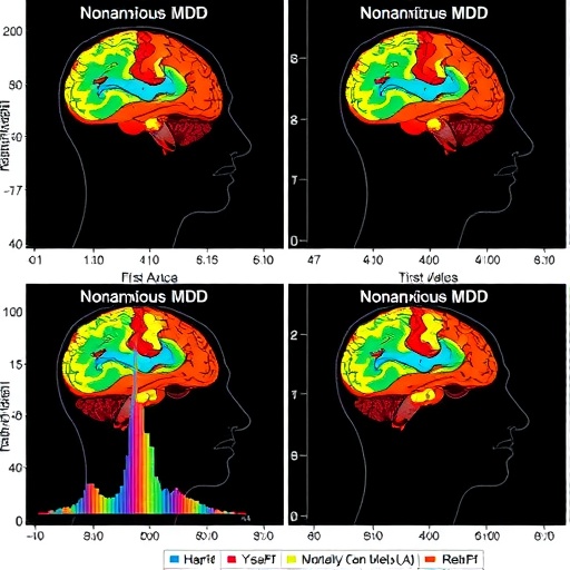
The researchers harnessed resting-state fMRI to measure regional homogeneity (ReHo)—a metric reflecting local synchronization of neuronal activity—providing a window into the intrinsic functional architecture of the brain. Subjects included 42 anxious MDD patients, 42 nonanxious MDD patients, and 45 healthy controls. Controlling for gray matter volume ensured that observed differences in ReHo values could be attributed to functional abnormalities rather than structural brain changes. This comprehensive approach allowed for nuanced comparisons aimed at identifying both shared and unique disruptions in brain activity.
Results revealed a striking convergence and divergence in regional brain dysfunctions between the two MDD subgroups. Both anxious and nonanxious patients exhibited decreased ReHo in several key brain areas, including the inferior temporal gyrus, precuneus, supplementary motor area, and putamen, underscoring a possible common neural signature of MDD. These regions are associated with cognitive processing, self-referential thinking, motor planning, and reward—functions often disrupted in depressive states. The observed hypoactivity suggests diminished spontaneous neuronal synchronization, which could underlie characteristic symptoms such as cognitive slowing and impaired motivation.
However, the distinction emerged with the nonanxious MDD group demonstrating additional reduced ReHo in the middle frontal gyrus, a region integral to executive function and emotion regulation. This unique alteration may hint at differential impairments in top-down cognitive control mechanisms, potentially accounting for variations in symptom profiles and treatment responsiveness. The supplementary motor area, inversely correlated with depression severity scores, highlights its role as a neural marker reflective of clinical state intensity.
Perhaps most notably, decreased ReHo in the right parahippocampal gyrus was found exclusively in anxious MDD patients when contrasted against their nonanxious counterparts. The parahippocampal region, intimately involved in memory encoding and emotional processing, has long been implicated in anxiety disorders. This selective hypoactivity could signify a neural correlate of heightened anxiety symptoms within depressive pathology, offering a promising target for diagnostic and therapeutic exploration.
To assess the diagnostic utility of these neuroimaging findings, the team employed receiver operating characteristic (ROC) curve analysis, demonstrating that altered activity in the right parahippocampal gyrus could effectively distinguish anxious from nonanxious depression. This represents a vital step toward the realization of objective biomarkers in psychiatry, which to date has largely relied on subjective clinical assessments. Such biomarkers could enable early identification, personalized treatment approaches, and more precise monitoring of therapeutic outcomes.
From a mechanistic perspective, the broader depression-related decreases in spontaneous brain activity found in this study reinforce existing evidence of disrupted intrinsic brain networks in MDD. The amplified regional hypoactivity in anxious patients suggests that comorbidity may reflect an additive or synergistic neural pathology rather than merely overlapping symptomatology. This distinction is critical as it advocates for tailored interventions addressing both depressive and anxious components simultaneously.
The implications of these findings extend beyond academic curiosity, offering practical insights that clinicians and researchers alike can employ. Better understanding the neurophysiological divergence between anxious and nonanxious MDD informs prognosis and therapeutic direction. It may guide clinicians to consider adjunct treatments targeting anxiety circuits in depression or to refine psychotherapeutic strategies based on differential brain activity profiles.
Moreover, this research exemplifies the power of advanced neuroimaging combined with stringent patient selection criteria, illuminating pathways toward more precise psychiatric diagnostics that transcend the limitations of symptom-based classification. Identifying reliable neurobiological markers is a critical milestone in achieving the holy grail of psychiatry: personalized medicine grounded in objective measurement rather than trial-and-error prescribing.
Future investigations are warranted to replicate these findings in larger cohorts and explore longitudinal changes across treatment courses. Integrating multimodal imaging techniques, genetic profiling, and electrophysiological measures will enrich our understanding of the complex brain-behavior relationships in depressive disorders with anxiety. These multifaceted approaches promise to unravel the dynamic interplay of neurocircuitry that underpins symptom heterogeneity, ultimately enhancing patient outcomes.
In conclusion, the study unravels critical differences in spontaneous brain activity between first-episode drug-naïve anxious and nonanxious MDD patients, highlighting the right parahippocampal gyrus as a potential neuroimaging biomarker for anxiety comorbidity in depression. By delineating both shared and unique regional dysfunctions, the research offers a refined neurobiological framework for understanding and managing major depressive disorder’s multifaceted clinical spectrum. This innovation marks a significant stride toward precision psychiatry, where neuroimaging can guide diagnosis and tailor interventions in what has traditionally been a challenging mental health landscape.
Subject of Research: Investigation of regional brain activity differences in first-episode drug-naïve anxious versus nonanxious major depressive disorder patients.
Article Title: Similarities and differences of regional brain activity between first-episode drug-naïve anxious and nonanxious MDD patients.
Article References:
Liu, G., Sun, Z., Zhou, C. et al. Similarities and differences of regional brain activity between first-episode drug-naïve anxious and nonanxious MDD patients. BMC Psychiatry 25, 1034 (2025). https://doi.org/10.1186/s12888-025-07513-9
Image Credits: AI Generated
