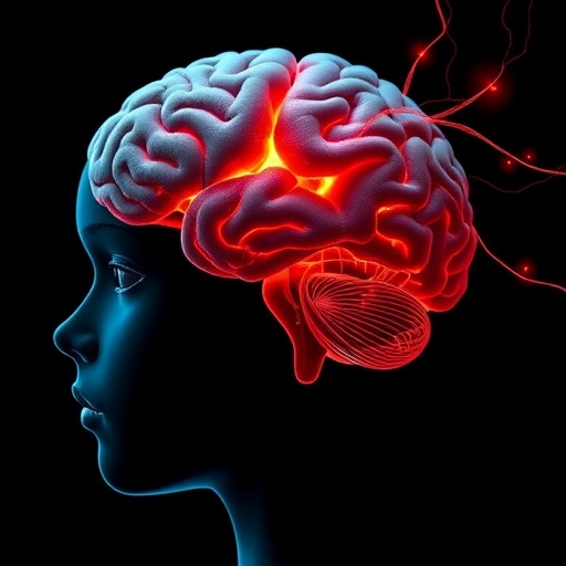
In recent years, the intricate relationship between brain connectivity and mental health has become a pivotal focus within neuroscience and psychiatry. Now, a groundbreaking study published in Translational Psychiatry sheds unprecedented light on how the connectivity between two critical brain regions, the amygdala and hippocampus, correlates with childhood depressive symptoms. This investigation delves deeper than ever before, parsing the nuanced interactions between subnuclei within these structures and revealing a complex network that underpins early depressive states in children. The research further explores how these neurobiological mechanisms intertwine with aspects of self-concept, offering a multifaceted perspective on depression’s developmental origins.
The amygdala and hippocampus are famed for their roles in emotion processing and memory formation, respectively. Traditionally, their functions are studied individually, but recent advances have allowed scientists to examine their connectivity with remarkable precision. The study employs cutting-edge neuroimaging techniques to examine the connectivity patterns between specific subnuclei of the amygdala and hippocampus. By moving beyond gross anatomical analyses, the authors have mapped how distinct subregions within these structures communicate and how such interactions vary with depressive symptomatology in childhood.
This subnuclear focus is pivotal because both the amygdala and hippocampus are not homogenous structures; instead, they consist of varied subfields with specialized functions and connectivity profiles. For instance, the basolateral nucleus of the amygdala is implicated in emotional evaluation, while the hippocampal CA1 and CA3 subfields are critical for different facets of memory processing. By delineating connectivity at such granular levels, the study reveals that depressive symptoms are associated with alterations in specific amygdala-hippocampus subnuclei pathways rather than broad regional dysfunction. This precision offers new targets for early interventions aimed at emotional regulation deficits seen in childhood depression.
Methodologically, the research integrates sophisticated imaging modalities with robust statistical modeling. Functional magnetic resonance imaging (fMRI) was utilized to capture resting-state connectivity, providing insights into the intrinsic communication between amygdalar and hippocampal subnuclei without task-induced activations. This is complemented by detailed symptom assessments conducted through validated psychometric instruments, ensuring that the neurobiological data correspond closely with behavioral and emotional phenotypes observed in the young participants. Advanced network analyses, such as graph theoretical metrics, were also employed to quantify the strength and efficiency of subnuclei connections, highlighting pathways that diverge in children exhibiting depressive tendencies.
One of the study’s most revelatory findings is the altered connectivity pattern between the central nucleus of the amygdala and the dentate gyrus of the hippocampus. These subregions are integral to modulating stress responses and memory encoding of emotional experiences. The diminished connectivity observed here suggests a neurobiological substrate for the dysregulated emotional processing and negative cognitive biases characteristic of childhood depression. Equally compelling is the hyperconnectivity found between the basolateral amygdalar nucleus and the CA3 hippocampal subfield, which may underlie heightened sensitivity to negative emotional stimuli, exacerbating depressive symptoms.
Crucially, the study does not stop at mapping connectivity changes; it examines how these neural alterations relate to children’s self-concept — a psychological construct encompassing self-esteem, self-worth, and self-identity. Using comprehensive assessments, the researchers established that children with disrupted amygdala-hippocampus subnuclei connectivity scores often concurrently reported impaired self-concept measures. This synergy between brain connectivity and self-perception heralds a more integrated model of childhood depression, suggesting that neural circuit abnormalities contribute directly to maladaptive self-related cognitions that perpetuate depressive states.
The implications of this research are vast, particularly in terms of early detection and personalized treatment. Understanding the subnuclear connectivity patterns that predispose children to depressive symptomatology enables clinicians to refine their diagnostic criteria. It also opens avenues for targeted therapies that aim to restore or compensate for specific circuit dysfunctions rather than employing a one-size-fits-all approach. For example, neurofeedback or neuromodulation techniques could potentially be tailored to normalize aberrant amygdala-hippocampal interactions, thereby improving therapeutic outcomes.
Moreover, these findings resonate with developmental neuroscience paradigms emphasizing the brain’s plasticity. Since childhood represents a period of profound neural remodeling, early disruptions in amygdala-hippocampus communication may set the stage for chronic mood disorders if left unaddressed. Therefore, this study underscores the urgency of identifying biomarkers of depression during early developmental windows, which could guide preventive strategies and mitigate long-term morbidity associated with mood disorders.
Technological progress was instrumental in facilitating the resolution needed for such subnuclear investigations. The deployment of ultra-high-field 7-Tesla MRI scanners allowed the researchers to visualize brain connectivity at an unprecedented spatial resolution. This advancement afforded them the capacity to differentiate between closely situated subnuclei, overcoming prior limitations imposed by conventional imaging. Additionally, the use of refined data preprocessing pipelines reduced noise and enhanced the reliability of connectivity measures, particularly important in pediatric neuroimaging studies prone to motion artifacts.
Importantly, the interdisciplinary synergy driving this research cannot be overstated. Neurologists, psychologists, and computational neuroscientists collaborated to weave together a richly detailed tapestry of neurobiological insight paired with psychological reality. Their integrative approach exemplifies the trend in modern neuroscience toward combining multimodal data to unravel complex brain-behavior relationships. This study stands as a testament to how collaborative frameworks can accelerate the translation of neuroscience findings into clinically meaningful knowledge.
Another layer of interest arises from the cultural and environmental context of the participants. The researchers accounted for variables such as socioeconomic status, family dynamics, and exposure to early-life stress, all known to influence brain development and depression risk. Controlling for these factors strengthened confidence that the reported neural connectivity alterations reflect intrinsic pathological processes rather than confounded epiphenomena. Such rigor enhances the generalizability of the findings across diverse populations, a critical consideration for future global mental health initiatives.
From a theoretical standpoint, these findings challenge simplistic models of depression that focus solely on monoaminergic dysfunction or regional brain volume changes. Instead, they align with network-based conceptualizations positing that mood disorders arise from disrupted communication within and between critical brain circuits. The subnuclear specificity introduced here refines this network model, suggesting that delicate balance and fine-tuning of connectivity at subregional scales are essential for emotional equilibrium, especially during formative years.
Furthermore, this study’s investigation into the interplay between brain connectivity and self-concept introduces novel psycho-neurological intersections. It raises intriguing questions about causality and directionality: does abnormal brain connectivity lead to distorted self-concept, or do negative self-perceptions influence neural communication patterns? Deciphering these pathways will be crucial for designing comprehensive therapeutic approaches that address both neural and cognitive dimensions of depression.
Future research inspired by these results will likely focus on longitudinal designs, tracking connectivity and self-concept over time to delineate trajectories leading to remission or exacerbation of depressive symptoms. Incorporating interventions aimed at modifying self-concept may also reveal how plastic these neural networks are and to what extent psychosocial variables modulate connectivity. Ultimately, such endeavors could revolutionize preventive mental health care by integrating neurobiological markers into routine pediatric assessments.
In sum, this pioneering study provides a transformative lens through which to view childhood depression, highlighting the importance of amygdala-hippocampus subnuclei connectivity and its relationship with self-concept. It punctuates the notion that mental health disorders emerge from complex interactions between brain circuits and psychological constructs, emphasizing precision medicine’s future potential. As neuroscience tools continue to evolve, unraveling these intricate connections will pave the way for more effective, individualized treatments that begin far earlier in life stages than previously possible.
Subject of Research: Amygdala-hippocampus connectivity in relation to childhood depressive symptoms and self-concept.
Article Title: Amygdala-hippocampus connectivity and childhood depressive symptoms: subnuclei insights and self-concept roles.
Article References:
Luo, L., Huang, P., Chan, S.Y. et al. Amygdala-hippocampus connectivity and childhood depressive symptoms: subnuclei insights and self-concept roles. Transl Psychiatry 15, 293 (2025). https://doi.org/10.1038/s41398-025-03524-y
Image Credits: AI Generated
