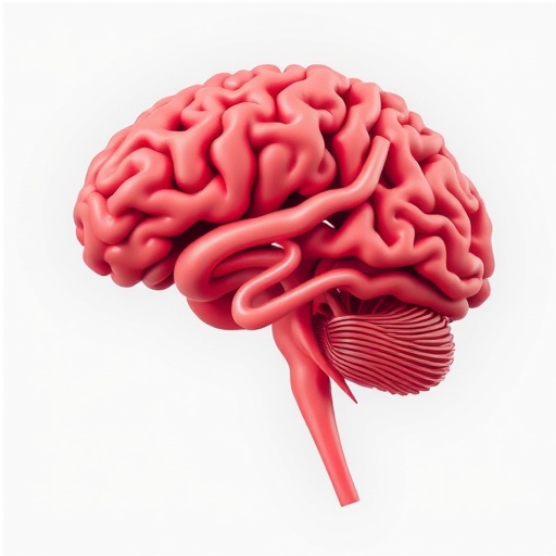
In the evolving landscape of mental health interventions, aerobic exercise (AE) has garnered significant attention as a promising non-pharmacological approach to alleviating depressive symptoms. Recent research published in BMC Psychiatry in 2025 delves into the neurobiological mechanisms underpinning this therapeutic modality by exploring the relationship between functional connectivity of the amygdala and symptom improvement in individuals with subthreshold depression (StD). This exploratory study leverages advanced neuroimaging techniques to elucidate how baseline brain connectivity patterns may predict responsiveness to AE, offering a window into personalized treatment strategies for depressive disorders.
Subthreshold depression, characterized by depressive symptoms that are clinically significant yet insufficient to meet full diagnostic criteria for major depressive disorder, poses a unique clinical challenge. The heterogeneity of symptom response to AE within this population underscores the necessity for biomarker identification that can forecast treatment outcomes. The amygdala, a critical brain region implicated in emotion regulation and mood disorders, presents a logical focal point for investigating such predictive markers due to its extensive connectivity within affective and cognitive neural circuits.
The study enrolled 43 participants diagnosed with StD who underwent a structured AE intervention designed to assess changes in depressive symptomatology. Pre- and post-intervention assessments were conducted using the Patient Health Questionnaire-9 (PHQ-9), a standardized clinical tool for quantifying depression severity. Participants were dichotomized into remitters and non-remitters based on their post-intervention PHQ-9 scores, enabling the examination of differential neural connectivity patterns relative to treatment efficacy.
Resting-state functional magnetic resonance imaging (rs-fMRI) served as the cornerstone of the neuroimaging methodology, capturing spontaneous brain activity and functional connectivity without the influence of task performance. By focusing on the amygdala’s connectivity with various cortical and subcortical structures, the research probed the neural substrates that might underlie symptom amelioration following AE. This approach capitalizes on the premise that intrinsic connectivity patterns can reveal latent neural circuit configurations associated with treatment responsiveness.
Analyses revealed compelling associations between baseline amygdala functional connectivity and depressive symptom outcomes post-exercise. Specifically, increased connectivity of the left amygdala with the right precuneus and bilateral middle frontal gyrus (MFG) was positively correlated with higher PHQ-9 scores after the intervention, indicating poorer symptom remission. Conversely, connectivity of the left amygdala with the left precuneus and left MFG exhibited negative correlations with symptom improvement, suggesting a nuanced relationship between neural circuit dynamics and therapeutic benefit.
Intriguingly, remitters demonstrated significantly reduced functional connectivity between the left amygdala and the left supplementary motor area (SMA) compared to non-remitters. This finding hints at the SMA’s potential role in modulating mood-related neural networks in the context of AE and points toward decreased amygdala-SMA coupling as a marker of positive treatment response. The SMA’s involvement in motor planning and cognitive control may interface with emotional regulation pathways, providing a plausible mechanistic substrate for observed effects.
Explorations of the right amygdala’s connectivity painted a slightly different picture. Enhanced connectivity between the right amygdala and regions including the left inferior parietal lobe (IPL), right middle temporal gyrus (MTG), left superior medial frontal gyrus (mSFG), and left MTG correlated with higher residual depressive symptoms post-intervention. However, these associations did not extend to symptom change metrics or group-level differences, underscoring possible lateralization effects in amygdala functional connectivity related to treatment outcomes.
The study further employed integrative analyses combining bilateral amygdala connectivity data with clinical variables, yielding robust classification accuracy (AUC = 0.93) within the sample for distinguishing remitters from non-remitters. This high predictive power underscores the practical potential of neuroimaging biomarkers in forecasting individual response to AE, a significant leap toward precision medicine paradigms in psychiatry. The ability to predict responders prior to intervention could optimize resource allocation and tailor treatment plans.
Despite the promising findings, the absence of significant group-by-time interactions in the connectivity patterns tempers the interpretation, suggesting that the observed functional connectivity differences were not dynamically altered by the AE intervention over time but rather reflected baseline neural states predictive of outcome. This nuance invites further longitudinal and interventional studies to unravel causality and temporal dynamics in neuroplasticity associated with exercise-based therapies.
The implications of these findings resonate beyond the immediate context of subthreshold depression. They highlight the intricate interplay between neurocircuitry and behavioral intervention efficacy, emphasizing the need to integrate neurobiological assessments into clinical practice. The amygdala’s connectivity profile emerges as a potential biomarker not only for predicting AE responsiveness but also for guiding adjunctive therapeutic strategies, including neuromodulation or cognitive-behavioral interventions.
While exploratory, this research marks a critical step in decoding the neural correlates of exercise-induced mood improvement. The deployment of rs-fMRI to reveal individual differences in brain connectivity furthers our understanding of depression’s neural architecture and its modulation by lifestyle factors. Future investigations expanding sample sizes and incorporating control conditions are essential to validate and extend these insights.
This novel perspective invigorates the discourse on exercise psychiatry, bridging neuroimaging advancements with clinical symptomatology. It encourages a paradigm shift towards leveraging functional brain metrics in the design and monitoring of interventions. As the mental health field grapples with treatment heterogeneity and accessibility challenges, such neurobiologically informed approaches could revolutionize care pathways, optimizing outcomes through personalized medicine.
In conclusion, the study underscores the pivotal role of amygdala-based functional connectivity in modulating depressive symptoms in response to aerobic exercise among individuals with subthreshold depression. By illuminating neural predictors of treatment response, this work paves the way for integrating neuroimaging biomarkers into clinical algorithms for depression management, promising more targeted and effective non-pharmacological interventions in mental health care.
Subject of Research: Functional connectivity of the amygdala as a neural predictor of response to aerobic exercise in subthreshold depression
Article Title: Amygdala functional connectivity and response to aerobic exercise in subthreshold depression-an exploratory fMRI study
Article References:
Huang, L., Zhang, W., Zhang, J. et al. Amygdala functional connectivity and response to aerobic exercise in subthreshold depression-an exploratory fMRI study. BMC Psychiatry 25, 1078 (2025). https://doi.org/10.1186/s12888-025-07535-3
Image Credits: AI Generated
DOI: 10.1186/s12888-025-07535-3 (Published 11 November 2025)


