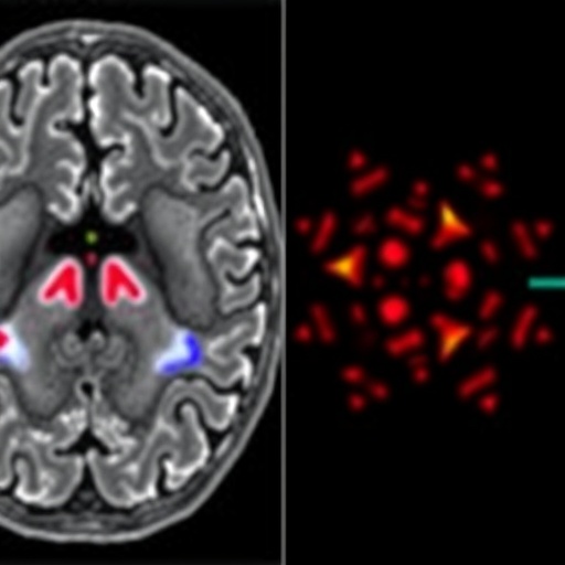
A groundbreaking artificial intelligence system developed by researchers at the University of Michigan is poised to revolutionize the field of neuroimaging by rapidly interpreting brain MRI scans and providing near-instantaneous diagnoses. This AI-powered model, dubbed Prima, demonstrated an extraordinary ability to detect a wide spectrum of neurological conditions with accuracy rates approaching 97.5 percent. Beyond diagnosis, Prima can also assess the urgency of each case, effectively prioritizing patients who need immediate intervention.
The innovative technology promises to alleviate the escalating pressures faced by healthcare systems worldwide, particularly the increasing demand for MRI scans which strains radiology departments and neurologists. Unlike traditional approaches that depend heavily on manual analysis of MRI data, Prima utilizes a vision language model (VLM) architecture, enabling it to simultaneously process multimodal inputs including images, videos, and textual clinical data. This approach mirrors the comprehensive assessment methods used by expert radiologists.
Prima was trained on an unprecedentedly vast dataset comprising over 200,000 MRI studies encompassing 5.6 million imaging sequences, spanning decades of digitized radiology records from the University of Michigan Health system. This expansive training corpus, incorporating both imaging data and patients’ clinical histories alongside physicians’ indications for ordering scans, has allowed the system to develop a broad and nuanced understanding of neurological health, enhancing its diagnostic capabilities across more than 50 distinct radiologic diagnoses.
Through rigorous testing over 30,000 MRI scans collected during a full year, the model consistently outperformed existing state-of-the-art AI systems, not only in diagnostic accuracy but also in its ability to triage cases based on urgency. For critical neurological emergencies—such as brain hemorrhages or ischemic strokes—the system can autonomously flag cases and issue real-time alerts to relevant specialists, facilitating expedited clinical responses.
The capacity of Prima to assign the appropriate subspecialty expertise—whether to stroke neurologists, neurosurgeons, or neuro-oncologists—underscores its potential to streamline workflows in clinical environments. This automation enhances decision-making efficiency without compromising diagnostic precision, addressing a crucial bottleneck that often results from the limited availability of neuroradiology specialists, particularly in resource-constrained or rural healthcare settings.
Prima’s architecture as a vision language model is especially notable for its integration of multi-format data inputs. Unlike prior AI models limited to narrow tasks—such as lesion detection or dementia risk prediction—Prima embodies a holistic analytic paradigm. It assimilates imaging information in concert with patient history to construct a comprehensive clinical context, thus reflecting the multifaceted diagnostic process employed by human radiologists.
The demand for MRI studies, especially those focused on neurological disorders, surpasses the capacity of current healthcare infrastructures, accentuating risks such as diagnostic delays and human errors. The advent of AI systems like Prima heralds a transformative advance, improving access to timely and accurate neuroimaging interpretations across diverse healthcare environments.
Looking forward, the researchers intend to augment Prima’s capabilities by incorporating more granular patient data drawn from electronic health records. This integration will further refine diagnostic precision, enabling personalized assessments that more effectively guide treatment decisions. Such advancements aim to bridge the gap between radiological imaging and patient-specific clinical realities.
The broader implications of Prima extend beyond neuroimaging. Todd Hollon, M.D., the study’s senior author and neurosurgeon, envisions the technology adapting to a range of medical imaging modalities, including mammography, chest radiography, and ultrasound diagnostics. Its analogy as a “ChatGPT for medical imaging” reflects its versatility as an AI co-pilot, assisting clinicians by generating diagnostic insights and recommendations that augment human expertise.
This pioneering work is the culmination of multidisciplinary collaboration among neurosurgeons, data scientists, radiologists, and computer engineers at the University of Michigan, supported by prominent funding entities such as the National Institute of Neurological Disorders and Stroke, the Chan Zuckerberg Initiative, and several philanthropic foundations. The results substantiated in the peer-reviewed journal Nature Biomedical Engineering herald a paradigm shift in how health systems may harness AI to tackle the challenges of modern clinical diagnostics.
Despite its promising performance, Prima remains in the early phases of clinical evaluation. Continued validation and real-world implementation studies will be critical to ascertain its safety, efficacy, and integration within healthcare workflows. Moreover, engagement with healthcare providers, policymakers, and regulatory bodies is underway to establish frameworks ensuring ethical and effective deployment of AI in medical imaging.
Ultimately, Prima exemplifies the transformative potential of artificial intelligence in healthcare, offering a scalable solution to the ever-growing diagnostic demands in neurology. By combining comprehensive data integration, rapid processing, and actionable clinical outputs, this AI-driven innovation stands to significantly improve patient outcomes, reduce diagnostic errors, and streamline neuroimaging practices across diverse care settings worldwide.
Subject of Research: People
Article Title: Learning neuroimaging models from health system-scale data
News Publication Date: 6-Feb-2026
Web References:
- https://www.nature.com/articles/s41551-025-01608-0
- http://dx.doi.org/10.1038/s41551-025-01608-0
References: “Learning neuroimaging models from health system-scale data,” Nature Biomedical Engineering, DOI: 10.1038/s41551-025-01608-0
Keywords: Artificial intelligence, Imaging, Medical imaging, Neurological disorders, Neurology, Neurosurgery
