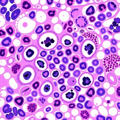
In the realm of medical diagnostics, histopathology stands as a cornerstone technique, providing critical insights into tissue morphology and disease progression. However, traditional histopathological workflows are notoriously time-consuming, labor-intensive, and prone to technical artifacts. A recent groundbreaking study led by Ma, J., Li, W., Li, J., and colleagues published in Nature Communications in 2026 promises to revolutionize this domain by integrating generative artificial intelligence (AI) into the virtual staining process. This development not only accelerates histopathological analysis but also offers unprecedented resilience to common issues like slide misalignment, a notorious hurdle in digital pathology.
Histopathological staining is essential for visualizing cellular and tissue structures, enabling pathologists to distinguish between healthy and diseased tissues. The conventional process involves physically applying chemical dyes to thin sections of tissue, a procedure that can take hours or even days from sample preparation to image acquisition. Delays in staining inevitably slow down diagnostic timelines, adversely affecting patient outcomes, especially in time-sensitive diseases like cancer. The novel approach put forward by Ma et al. leverages generative AI models to simulate staining digitally, abolishing the need for physical dyes and dramatically compressing analysis timeframes.
The core innovation lies in a generative adversarial network (GAN)-inspired architecture optimized to produce virtual stains from unstained tissue images captured via light microscopy. Unlike prior attempts that suffered from sensitivity to misalignment between images of the same tissue section before and after staining, this pioneering technique exhibits remarkable tolerance to such discrepancies. Slide misalignment, often caused by mechanical shifts during sample preparation or imaging, typically degrades the accuracy of virtual staining algorithms, affecting downstream diagnostic interpretations. Ma and colleagues tackled this challenge head-on by embedding misalignment-resistant features directly into their generative AI framework.
Their AI model was trained on a massive dataset comprising paired unstained and stained tissue sections from diverse histological contexts, including oncology, inflammatory diseases, and organ pathology. By learning the complex morphological correlations between unlabeled and stained tissues without relying on pixel-perfect alignment, the system demonstrates extraordinary robustness. This allows pathologists to generate reliable virtual stains even when physical slide alignment is suboptimal—a capability that could redefine quality control standards in digital pathology laboratories.
The implications of this technology on workflow efficiency are monumental. With conventional staining pipelines requiring significant manual intervention and reagent costs, virtual staining using generative AI could dramatically cut both labor expenses and consumable usage. Moreover, clinical turnaround times could be shortened from several days to mere hours or even minutes. Given the pressure hospitals face to deliver rapid, accurate diagnoses amidst increasing caseloads, this advancement introduces a scalable solution with the potential to alleviate bottlenecks in histopathology services globally.
Technically, the researchers innovated by incorporating spatial transformation modules and learned attention mechanisms within the GAN framework. These components enable the AI to internally correct for geometric distortions caused by tissue deformation or slide positioning errors. Consequently, the output virtual stains preserve intricate cellular details such as nuclear morphology, cytoplasmic texture, and extracellular matrix patterns—hallmarks critical for precise histopathological interpretation. This fidelity ensures that virtual staining is not just a qualitative image transformation but a quantitatively trustworthy surrogate for conventional staining methods.
Additionally, this approach harmonizes seamlessly with existing digital pathology infrastructures. Digital slides acquired via whole-slide imaging (WSI) scanners can be input directly into the AI model, producing virtual stains without additional hardware or procedural adjustments. This compatibility paves the way for rapid adoption in pathology labs, mitigating the need for extensive retraining or equipment overhaul. Beyond efficiency gains, the AI-driven process also minimizes human error and inter-operator variability, enhancing diagnostic consistency across institutions and geographic locations.
The study also delves into the scalability of the model across various staining protocols, demonstrating the technique’s flexibility. While hematoxylin and eosin (H&E) remain the gold standard in most tissue assessments, there are instances requiring specialized stains to highlight particular cell types or extracellular components. Ma et al.’s model architecture can be trained with diverse staining datasets, enabling virtual replication of complex stains such as immunohistochemistry or special stains like PAS or Masson’s trichrome. This adaptability signifies a future where pathologists may access a virtual pallet of staining techniques instantly, tailored to clinical needs.
The accelerated staining workflows enabled by this generative AI approach are particularly beneficial for large-scale research studies and biobanking operations. High-throughput histological assessments traditionally necessitate extensive reagent stockpiling and prolonged sample batching. Virtual staining could permit real-time analysis, allowing researchers to make time-sensitive decisions or correlations during experimental protocols. Moreover, the digital nature of the stained images facilitates integration with advanced image analysis and machine learning tools for automated feature extraction, potentially supercharging biomarker discovery pipelines.
Crucially, the researchers emphasize the interpretability and validation of the AI outputs. They employed rigorous quantitative metrics, including structural similarity indices and expert pathologist concordance, to benchmark virtual stains against their physical counterparts. Their validation pipelines ensure the AI-generated images are not mere aesthetic reproductions but adhere to diagnostic criteria essential for clinical decision-making. This level of scrutiny is indispensable to gain regulatory approvals and physician trust in deploying generative AI in routine pathology workflows.
Ethical considerations surrounding AI in medicine also find a thoughtful discussion within the study. By reducing reliance on chemical reagents, the approach contributes to eco-friendly pathology practices. Furthermore, the digitization of staining fosters remote diagnostics and telepathology, increasing healthcare access for underserved populations. Nevertheless, the authors caution against sole dependence on algorithmic outputs without expert oversight, advocating for a hybrid paradigm where AI serves as a powerful adjunct rather than a replacement for human expertise.
As this field moves forward, integration with multimodal imaging systems leveraging fluorescent or multiphoton microscopy could further augment the virtual staining capabilities. The adaptability of generative AI to different tissue imaging modalities opens avenues for exploring subcellular and molecular level virtual stains that might one day surpass the limitations of traditional histochemical dyes. Such innovation could unlock richer diagnostic insights, pushing the frontier of personalized medicine and targeted therapies.
The intersection of AI and histopathology epitomized by this study embodies a broader trend toward digitization and computational augmentation in medicine. The misalignment-resistant virtual staining model by Ma et al. signals a transformative leap, marrying cutting-edge AI techniques with pragmatic clinical needs. As this technology matures and gains regulatory acceptance, it could democratize access to high-quality pathology diagnostics, reduce operational costs, and expedite therapeutic interventions—ultimately reshaping the landscape of diagnostic pathology in profound and lasting ways.
Subject of Research:
Generative AI applications in histopathology for misalignment-resistant virtual staining techniques to accelerate tissue diagnostics.
Article Title:
Generative AI for misalignment-resistant virtual staining to accelerate histopathology workflows.
Article References:
Ma, J., Li, W., Li, J. et al. Generative AI for misalignment-resistant virtual staining to accelerate histopathology workflows. Nature Communications (2026). https://doi.org/10.1038/s41467-026-71038-2
Image Credits:
AI Generated
DOI:
https://doi.org/10.1038/s41467-026-71038-2
