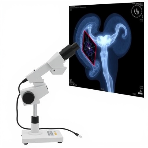
In a groundbreaking convergence of artificial intelligence and medical diagnostics, researchers have unveiled an innovative approach to cervical precancerous screening designed specifically for high-risk populations residing in resource-limited regions. This pioneering work leverages an AI-assisted compact microscope system, promising a transformative shift in the early detection and management of cervical cancer, a disease that remains one of the leading causes of cancer-related mortality among women worldwide, particularly in low-resource settings. The new technology represents a significant stride toward overcoming persistent barriers in healthcare accessibility, diagnostic accuracy, and cost-efficiency that have long hindered effective cancer screening and prevention strategies.
Cervical cancer screening programs have historically faced numerous challenges, especially in under-resourced areas. Conventional methods typically require expensive laboratory equipment, highly trained cytopathologists, and well-established healthcare infrastructure, all of which are often lacking in regions with the greatest need. The AI-driven compact microscope system, developed through a multidisciplinary collaboration, sidesteps these obstacles by integrating state-of-the-art artificial intelligence algorithms directly into a portable, user-friendly imaging platform. This democratizes access to vital cervical screening services, potentially saving thousands of lives through earlier intervention.
Central to this new diagnostic tool is an advanced AI model trained on extensive datasets of cervical cytology images, enabling the system to accurately identify morphological changes indicative of precancerous lesions. Unlike traditional manual screening methods, which are labor-intensive and prone to human error, the AI model offers consistent and rapid analysis, highlighting suspicious cells with high sensitivity and specificity. This capability is particularly crucial in high-risk populations where timely detection and treatment are often hampered by logistical and financial constraints.
The compact microscope component of the system is a marvel of engineering, combining affordability with high-resolution imaging capabilities. Its design prioritizes portability and ease of use, facilitating deployment in a wide range of clinical settings—from rural health clinics to mobile medical units. The light-weight and robust construction ensure that the device can operate efficiently despite environmental challenges common in resource-limited areas, such as unstable power supplies and inadequate laboratory facilities.
One of the most remarkable aspects of this innovation is the seamless integration of hardware and software components. The AI software runs locally on an embedded computing platform without necessitating cloud connectivity, addressing data privacy concerns and minimizing reliance on internet infrastructure, which is often unreliable in target deployment areas. This autonomy also accelerates diagnostic turnaround times, enabling healthcare workers to provide immediate counseling and referral decisions during patient visits.
The training process for the AI algorithm involved sophisticated image augmentation and annotation techniques to capture the wide variability of cellular features seen in diverse populations. By incorporating data from multiple demographic groups and geographic regions, the model achieves robust generalizability and avoids biases that could undermine diagnostic fairness. Regular updates and retraining protocols ensure that the system adapts continuously to emerging cytological patterns and maintains peak performance.
In pilot clinical trials conducted across several rural sites, the AI-assisted screening system demonstrated sensitivity and specificity rates comparable to those of expert cytopathologists. Notably, the reduction in false negatives empowers clinicians to catch early transformations that might otherwise progress undetected, while the decrease in false positives prevents unnecessary anxiety and invasive follow-ups. Patient feedback has been overwhelmingly positive, with many expressing appreciation for the expedited process and localized service delivery.
The impact of this technology extends beyond individual patient care, offering substantial public health benefits by enabling large-scale screening programs at a fraction of the traditional cost. Health ministries and non-governmental organizations are poised to deploy these systems in community-based initiatives, screening tens of thousands of women who previously lacked access to regular cervical cancer surveillance. This shift could dramatically reduce disease burden and associated economic costs in vulnerable populations.
Moreover, the AI-assisted platform fosters task-shifting opportunities, empowering mid-level healthcare providers and technicians to conduct reliable screenings without requiring the constant presence of highly specialized personnel. This reallocation of human resources alleviates workforce shortages and enhances the sustainability of cervical cancer control programs. Training modules accompanying the device ensure that end-users can efficiently operate the system and interpret results within existing care pathways.
The researchers emphasize that the technology’s scalability and adaptability signify an important advance in global health equity. While initially focused on cervical cytology, the underlying AI framework and compact imaging design hold potential applications in diagnosing other cytological and histological conditions, broadening the scope of affordable diagnostics for marginalized communities. Continuing collaborations between engineers, clinicians, and public health experts will be vital to realize this vision fully.
Challenges remain, including regulatory approvals, supply chain logistics, and integration with national health information systems. However, the early successes and promising data underscore the feasibility of AI-assisted cervical screening as a viable and impactful intervention. Ongoing real-world evaluations and cost-effectiveness analyses will further elucidate pathways for widespread adoption and sustained operation.
In an era increasingly defined by the synergy of artificial intelligence and medicine, this innovation stands out as a beacon of hope for millions of women at risk of cervical cancer. By delivering sophisticated diagnostic capabilities to the doorstep of those who need them most, the AI-assisted compact microscope system epitomizes the future of equitable, precision healthcare. As this technology moves closer to global implementation, it heralds a new chapter in the fight against preventable cancers, driven by ingenuity, collaboration, and a commitment to saving lives.
The study was published in Nature Communications and highlights a blueprint for harnessing AI and compact instrumentation to revolutionize disease screening in resource-limited environments. It offers a proof of concept for how technological advances can be repurposed to tackle some of the most persistent public health challenges, simultaneously addressing issues of affordability, accessibility, and clinical reliability.
This promising development invites further research into expanding AI diagnostic tools across a spectrum of diseases that disproportionately affect underserved populations. The lessons learned from this project underscore the critical importance of context-specific solutions that respect local constraints while leveraging cutting-edge innovation. Ultimately, the fusion of AI and compact imaging heralds a more just and effective era of healthcare delivery worldwide.
Subject of Research: AI-assisted cervical cytology precancerous screening in high-risk populations within resource-limited regions using a compact microscope.
Article Title: AI-assisted cervical cytology precancerous screening for high-risk population in resource-limited regions using a compact microscope.
Article References:
Bai, J., Li, N., Ye, H. et al. AI-assisted cervical cytology precancerous screening for high-risk population in resource-limited regions using a compact microscope. Nat Commun 16, 7429 (2025). https://doi.org/10.1038/s41467-025-62589-x
Image Credits: AI Generated


