In a groundbreaking study set to reshape gross anatomy education, researchers have meticulously compared traditional physical prosections with cutting-edge virtual 3D scanned models to gauge their effectiveness as teaching tools for the external human heart. This innovative research, involving twenty-nine incoming medical students, meticulously assessed both knowledge acquisition and learner confidence, providing robust data that challenges long-held assumptions about anatomical education.
The participants were systematically divided into two cohorts: one group engaged with conventional physical prosections, a tactile and time-honored method of learning human anatomy, while the other explored virtual 3D scanned prosections, employing advanced imaging technology to simulate real anatomical structures. This approach allowed researchers to directly contrast the educational outcomes derived from tactile, hands-on experiences and those facilitated through state-of-the-art digital technology.
Pre- and post-instruction assessments revealed that both groups exhibited statistically significant improvement in examination scores, with no discernible difference between the physical and virtual cohorts. These findings robustly demonstrate that virtual 3D scanned models can achieve comparable educational efficacy to physical prosections in teaching complex anatomical structures, specifically the external morphology of the heart. This equivalence in learning outcomes opens new horizons for digital anatomy education.
Beyond objective performance metrics, the study delved into subjective student perceptions, uncovering intriguing insights into learner confidence and acceptance of virtual models. Students exposed to the 3D virtual prosections reported enhanced confidence in their ability to assimilate anatomical knowledge using digital tools, highlighting the potential of immersive technology to engage and empower modern learners accustomed to digital interfaces.
Notably, despite recognizing the effectiveness and preparatory value of virtual 3D scans for dissection labs and examinations, all students unanimously agreed that digital replicas could not substitute the rich, immersive experience provided by hands-on dissection laboratories. The study underscores that physical dissection affords irreplaceable tactile feedback and emotional context critical for fostering respect for the human body and encouraging professional growth.
The unique tactile sensations experienced during physical dissection—which engage multiple sensory modalities—provide learners with kinesthetic memory and nuanced understanding unattainable via virtual manipulation. Such interactions facilitate deeper cognitive imprinting and ethical appreciation of human anatomy, elements considered essential for cultivating competent and empathetic future medical professionals.
This pivotal research also emphasizes that virtual 3D scanned prosections serve optimally as complementary educational resources rather than wholesale replacements for traditional dissection. Their integration into curricula can reinforce learning, provide flexible access to anatomical content, and augment preparation for diagnostic imaging interpretations and surgical simulations.
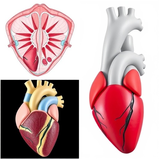
The technological foundation underpinning virtual prosection utilizes high-resolution 3D scanning and rendering techniques to create accurate, manipulatable digital surrogates of anatomical specimens. These models afford dynamic interactions—such as rotation, zooming, sectional slicing, and layer isolation—granting learners unprecedented control and visualization capabilities beyond the static physical specimen.
Furthermore, the deployment of virtual models aligns with increasing calls for digitization and remote learning solutions within medical education, especially highlighted by recent global disruptions limiting laboratory access. This adaptability signifies a critical step toward resilient, scalable, and inclusive teaching methodologies adaptable to diverse learning environments.
Nevertheless, experts caution that exclusive reliance on virtual anatomy risks depriving students of essential experiential learning that cultivates empathy, ethical sensibility, and manual dexterity—qualities foundational to medical professionalism. Optimal educational strategies should thus blend physical and virtual modalities to harness synergistic benefits of both.
The published work titled “A Practical Examination and Feedback Survey Evaluating Learners Taught Using Physical Prosections vs. 3D Models of Prosections of the External Heart” appeared in the eminent journal Frontiers of Digital Education on June 26, 2025, offering a timely and rigorous contribution to the evolving discourse on digital transformations in medical training.
In sum, this study marks a significant milestone in anatomical pedagogy, evidencing that virtual 3D scanned prosections are not only effective but also well-received by learners, yet affirming the irreplaceable value of physical dissection. As educational institutions worldwide strive to modernize anatomy instruction, these insights provide critical guidance to balance technological innovation with experiential tradition.
The implications extend beyond gross anatomy to specialties leveraging digital visualization, such as radiology and surgery, where precise spatial understanding is paramount. By validating digital prosections as effective learning tools, this research invites a broader reimagining of medical curricula optimized to future-proof healthcare education.
Continued interdisciplinary collaboration between anatomists, educators, and technologists will be crucial to refining virtual anatomy tools, enhancing their realism, interactivity, and pedagogical utility. Future studies may explore integration with augmented reality, haptic feedback systems, and adaptive learning platforms to further advance the digital anatomy frontier.
Ultimately, cultivating proficient physicians necessitates a harmonious fusion of tactile experience and digital innovation—a duality exemplified by this study’s findings. Medical educators are now better equipped to design anatomically rigorous, engaging, and accessible learning experiences that harness the best of both worlds.
Article Title:
A Practical Examination and Feedback Survey Evaluating Learners Taught Using Physical Prosections vs. 3D Models of Prosections of the External Heart
News Publication Date:
June 26, 2025
Web References:
http://dx.doi.org/10.1007/s44366-025-0064-9
References:
Stephanie A. Waldman, Zane Sejdiu, Shannon M. O’Hara, Jed S. Shumsky, Caitlin A. Howe. A Practical Examination and Feedback Survey Evaluating Learners Taught Using Physical Prosections vs. 3D Models of Prosections of the External Heart. Frontiers of Digital Education, 2025, 2(3): 27
Image Credits:
Stephanie A. Waldman, Zane Sejdiu, Shannon M. O’Hara, Jed S. Shumsky, Caitlin A. Howe
Keywords:
Information science, Applied sciences and engineering
