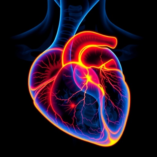
A groundbreaking advancement in molecular imaging technology promises to transform the clinical landscape for patients recovering from acute myocardial infarction (AMI), commonly known as a heart attack. Researchers have developed an innovative Positron Emission Tomography (PET) imaging technique that targets the cellular protein CXCR4, a critical mediator in the inflammatory response following cardiac injury. This novel approach enables clinicians to identify individuals at increased risk of adverse heart remodeling and heart failure, thereby providing a crucial window for timely therapeutic intervention.
Heart attacks represent a leading cause of morbidity and mortality worldwide, claiming hundreds of thousands of lives annually. The pathological aftermath of AMI involves a complex interplay of tissue injury, inflammation, and healing processes. A key challenge in cardiology has been the ability to predict which patients will experience functional cardiac recovery versus those who will develop progressive heart failure and deteriorating cardiac function. Traditional imaging modalities mainly quantify irreversible myocardial damage but fall short in capturing the dynamic inflammatory processes that dictate healing outcomes.
This newly developed CXCR4-targeted PET/CT imaging technology directly visualizes the spatial extent and magnitude of the inflammatory response by detecting upregulation of the CXCR4 receptor on inflammatory cells. CXCR4 plays a pivotal role in mediating leukocyte recruitment and retention at sites of injury, orchestrating the post-ischemic inflammatory cascade. By mapping CXCR4 expression shortly after AMI, researchers can glean mechanistic insights into the ongoing immune activity within the myocardium that profoundly influences remodeling and functional recovery.
In a comprehensive clinical study involving 49 patients who suffered an acute myocardial infarction, investigators employed a multimodal imaging protocol integrating CXCR4 PET/CT with myocardial perfusion imaging (MPI) and cardiac magnetic resonance imaging (MRI). Imaging was performed within the first week post-infarction, with follow-up MRI assessments approximately eight months later in 40 patients, allowing correlation between early inflammatory signals and long-term cardiac outcomes. This integrative approach represents a paradigm shift in cardiology diagnostics, combining molecular, functional, and structural data.
The study unveiled that CXCR4 expression extends beyond the infarct core into the border zone myocardium, areas adjacent to the primary damaged tissue previously underappreciated by conventional imaging methods. Importantly, elevated CXCR4 PET signal strongly correlated with subsequent left ventricular dysfunction, suggesting that protracted or excessive inflammation detected via CXCR4 imaging directly predicts detrimental cardiac remodeling trajectories. These findings establish a critical link between molecular inflammatory activity and future cardiac health.
Conventional diagnostic modalities such as MPI and cardiac MRI primarily delineate the scope of irreversible myocardial injury, capturing infarct size and scar formation but lacking the ability to dynamically assess the inflammatory milieu that guides healing. By incorporating CXCR4-targeted PET imaging, clinicians gain unprecedented access to the inflammatory microenvironment in vivo. This capability allows the identification of patients exhibiting pronounced or sustained inflammation, who may benefit from tailored anti-inflammatory or reparative treatments aimed at modulating post-infarction remodeling.
Dr. Johanna Diekmann, senior physician at the Department of Nuclear Medicine, Hannover Medical School, emphasized the clinical potential of this approach: “Our imaging strategy enables us to pinpoint those patients with excessive inflammatory response early after myocardial infarction. This information is invaluable in guiding personalized treatment plans that can alter the course of disease progression and improve long-term outcomes.” The prospect of image-guided therapeutic decision-making heralds a new era of precision cardiology.
Moreover, the molecular imaging technique opens avenues for monitoring the efficacy of novel therapeutics that target inflammation and tissue repair pathways. By serially performing CXCR4 PET scans, clinicians may track changes in inflammatory status and adjust interventions accordingly. This could significantly enhance the ability to optimize therapy and mitigate adverse remodeling before irreversible damage ensues. Ultimately, it positions nuclear medicine as an integral part of targeted cardiac care and regenerative medicine strategies.
The implications of these findings extend beyond clinical application to deepen scientific understanding of the inflammatory mechanisms underlying cardiac repair. The demonstration that CXCR4-mediated inflammation is spatially extensive and functionally significant reshapes our conceptual models of post-ischemic myocardial biology. It underscores the necessity of incorporating molecular inflammatory biomarkers into the standard diagnostic and prognostic arsenal for cardiovascular diseases.
The research team at Hannover Medical School, involving nuclear medicine specialists and cardiologists, highlights the power of interdisciplinary collaboration in tackling complex medical challenges. By leveraging advances in molecular imaging technology alongside state-of-the-art cardiac MRI and perfusion imaging, they provide a comprehensive snapshot of myocardial health that integrates cellular, structural, and functional perspectives.
In summary, CXCR4-targeted PET/CT represents a transformative breakthrough for post-myocardial infarction care. This precision imaging tool reveals the intricacies of myocardial inflammation and enables the early identification of patients prone to maladaptive remodeling and heart failure. The fusion of molecular imaging with conventional diagnostic modalities offers a robust framework for future research, personalized medicine, and improved clinical outcomes.
As this technology progresses through clinical validation and potential regulatory approval, it holds the promise of reshaping the management of millions of patients worldwide who suffer heart attacks annually. By furnishing timely, actionable insights into the inflammatory processes that govern healing, CXCR4 imaging paves the way toward a new frontier in cardiovascular diagnostics and therapy.
Subject of Research: Visualizing post-infarction inflammation using CXCR4-targeted PET imaging to predict cardiac functional recovery.
Article Title: CXCR4 PET/CT Predicts Left Ventricular Recovery 8 Months After Acute Myocardial Infarction
News Publication Date: October 21, 2025
Web References:
– https://doi.org/10.2967/jnumed.125.270807
– https://jnm.snmjournals.org/
References:
Diekmann, J., Hess, A., Ross, T.L., Thackeray, J.T., Bengel, F.M., Konig, T., Zwadlo, C., Schafer, A., Bauersachs, J. “CXCR4 PET/CT Predicts Left Ventricular Recovery 8 Months After Acute Myocardial Infarction.” Journal of Nuclear Medicine, 2025.
Image Credits: Image created by Johanna Diekmann, MD, senior physician at the Department of Nuclear Medicine, Hannover Medical School, Germany.
Keywords: Molecular imaging, Positron emission tomography, Medical imaging, Acute myocardial infarction, Cardiovascular disorders, Inflammation, CXCR4, Left ventricular remodeling, Cardiac MRI, Myocardial perfusion imaging, Precision medicine, Heart failure
