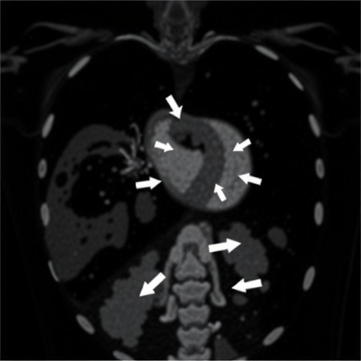
In the realm of neonatal medicine, congenital diaphragmatic hernia (CDH) remains one of the most challenging and complex conditions, often dictating survival outcomes within the fragile first hours and days of life. A new study, emerging from the collaboration of leading pediatric researchers, sheds unprecedented light on the early echocardiographic characteristics of CDH neonates and their critical role in predicting survival chances and the need for advanced interventions such as extracorporeal life support (ECLS). This research not only provides vital insight into the cardiac adaptations in these vulnerable infants but also paves the way for precision medicine approaches tailored to the unique physiological challenges posed by CDH.
CDH is a developmental anomaly characterized by an incomplete formation of the diaphragm, allowing abdominal organs to herniate into the thoracic cavity. This anatomical disruption severely compromises lung development, leading to pulmonary hypoplasia and pulmonary hypertension. While surgical repair is the definitive treatment, the timing and approach are intricately connected to the extent of pulmonary and cardiovascular compromise the neonate endures immediately postpartum. Until now, much of the prognostication rested on clinical observations and general assessments of lung function, but this study pivots the focus onto the subtle, yet critically informative, echocardiographic findings evaluated right after birth.
Echocardiography, a non-invasive ultrasound imaging technique, offers a real-time window into the heart’s structural and functional status. In the context of CDH, echocardiographic parameters can unmask the hemodynamic repercussions of pulmonary hypertension and cardiac remodeling driven by the distorted thoracic anatomy. The new research meticulously analyzed a cohort of neonates, systematically comparing echocardiographic features between those with varying laterality of the hernia—left versus right—and differing sizes of the diaphragmatic defect, thereby delineating nuanced cardiovascular phenotypes associated with each subgroup.
One of the pivotal contributions of this study is the identification of early echocardiographic markers associated with survival. Neonates exhibiting less pronounced right ventricular dysfunction and more favorable pulmonary artery pressures shortly after birth were noted to have significantly higher survival rates. This finding underscores the role of cardiac function as a pivotal determinant in the clinical trajectory of CDH infants, heralding the potential for integrating echocardiographic assessments into early risk stratification models that can guide both clinical decision-making and parental counseling.
Further, the study illuminates differential outcomes based on hernia laterality. Left-sided CDH, traditionally more common, showed distinct echocardiographic patterns compared to right-sided defects, influencing both survival probabilities and the likelihood of requiring extracorporeal life support. The cardiac alterations in right-sided CDH were often more severe, aligning with the worse prognoses observed in this group. This critical insight enhances our understanding of why not all CDH cases are created equal and accentuates the need for individualized therapeutic approaches.
Extracorporeal life support, a form of mechanical circulatory and respiratory support used when conventional therapies fail, remains a double-edged sword in CDH management. While it can be lifesaving, it is associated with significant risks and resource intensity. The ability to predict early which neonates are likely to require ECLS based on echocardiographic parameters represents a game changer. It promises a future where timely intervention can be orchestrated with precision, potentially mitigating adverse outcomes and optimizing resource allocation in neonatal intensive care units.
The investigative team employed advanced echocardiographic techniques, measuring variables such as right ventricular fractional area change, tricuspid annular plane systolic excursion (TAPSE), and pulmonary artery acceleration time. These precise metrics allowed a granular assessment of ventricular performance and pulmonary vascular resistance, crucial elements in the CDH physiopathology puzzle. Their comprehensive protocol, performed within hours of birth, provides a replicable framework for neonatal centers worldwide aiming to refine CDH prognostication.
Particularly intriguing was the analysis of defect size and its correlation with cardiac function and survival. Larger defects, often linked with greater pulmonary hypoplasia, demonstrated more pronounced cardiac strain and elevated pulmonary pressures, correlating with poorer outcomes. This association validates the anatomical basis of cardiac compromise in CDH and reinforces the multidimensional nature of risk factors, where anatomical severity intertwines with functional cardiac impairment to determine clinical fate.
The researchers also delved into the dynamic interplay between ventricular interdependence and septal morphology, observed through echocardiographic imaging. Altered septal curvature and interventricular septal shifts were recurrent in severe cases, reflecting the pathophysiological strain imposed by high pulmonary pressures. These insights into ventricular geometry alterations illuminate substrates for future targeted therapies aimed at ameliorating cardiac loading conditions in CDH neonates.
Interestingly, the temporal evolution of echocardiographic parameters was documented, revealing that early postnatal cardiac function indicators could predict subsequent clinical deterioration or recovery trajectories. This longitudinal perspective offers clinicians an invaluable monitoring tool, enabling the early identification of infants at risk of rapid decline who might benefit from escalated care or innovative therapeutic interventions.
The study also ventures into the potential integration of echocardiographic data with emerging biomarkers and genetic profiles, envisioning a holistic precision medicine model for CDH management. Such a multidisciplinary approach aligns perfectly with modern trends in neonatal care, where multifaceted data converge to inform personalized treatment paradigms, moving beyond a one-size-fits-all approach.
Implications of these findings ripple beyond immediate clinical practice. They inspire a recalibration of neonatal resuscitation protocols for CDH babies, emphasizing echocardiographic monitoring as a critical component of initial stabilization. Strategies for early pharmacologic modulation of pulmonary vascular resistance might also be tailored according to echocardiographic risk profiles, potentially altering disease course before irreversible damage occurs.
Moreover, the study advocates for standardized echocardiographic assessment pathways incorporated into national and international CDH registries. Such harmonization could foster large-scale data collection and meta-analyses, accelerating knowledge accumulation and optimizing guideline development. Bridging the gap between bedside imaging and clinical outcomes transforms echocardiography from a diagnostic tool into a prognostic powerhouse in neonatal care.
The ramifications for parental counseling are profound. By elucidating early markers linked with survival and the necessity for invasive support modalities, healthcare providers can engage families with clearer, evidence-based prognoses. This fosters informed decision-making and psychological preparedness, vital components of family-centered neonatal care.
This research also raises provocative questions prompting future inquiries. Could echocardiographic-guided interventions during the immediate neonatal period attenuate cardiac dysfunction and improve survival? What is the influence of prenatal echocardiographic findings on postnatal outcomes? Addressing these questions will propel the field into new frontiers of integrated perinatal care.
In sum, this landmark study delivers a comprehensive, evidence-backed examination of early echocardiographic phenomena in CDH neonates, unraveling complex cardiac dynamics that anchor survival and therapeutic needs. It sets a new standard for research and clinical praxis, promising improved outcomes through refined diagnostics and targeted interventions. As CDH continues to challenge the resilience of neonates and their caregivers, these insights fuel hope for transforming the prognosis of this formidable congenital condition.
Subject of Research: Early Echocardiographic Characteristics in Neonates with Congenital Diaphragmatic Hernia and Their Impact on Survival and Need for Extracorporeal Life Support
Article Title: Early postnatal echocardiographic characteristics impact survival and extracorporeal life support in congenital diaphragmatic hernia
Article References:
Noh, C.Y., Danzer, E., Bhombal, S. et al. Early postnatal echocardiographic characteristics impact survival and extracorporeal life support in congenital diaphragmatic hernia. Pediatr Res (2025). https://doi.org/10.1038/s41390-025-04443-w
Image Credits: AI Generated


