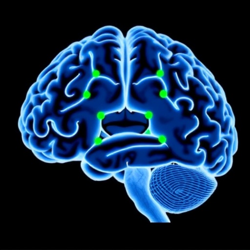
A revolutionary breakthrough in brain imaging has been achieved by researchers at the Mark and Mary Stevens Neuroimaging and Informatics Institute at the Keck School of Medicine of USC. This pioneering development has unveiled the potential to noninvasively visualize the volume changes in the brain’s tiny blood vessels—the microvasculature—as they pulse in rhythm with the heartbeat. This pulsatility, a rhythmic expansion and contraction within these smallest of vessels, may hold vital clues to understanding aging and neurological disorders such as Alzheimer’s disease.
Published recently in the prestigious journal Nature Cardiovascular Research, this study introduces a groundbreaking MRI technique that harnesses ultra-high field 7 Tesla (7T) magnetic resonance imaging to quantify cerebral microvascular volumetric pulsatility in unprecedented detail. By capturing dynamic changes occurring over the cardiac cycle, this approach is the first of its kind to measure microvascular pulsations in living humans safely and noninvasively, bombarding conventional limitations that confined prior investigations primarily to animal models.
At the core of this innovation lies the integration of two advanced MRI techniques: vascular space occupancy (VASO) imaging and arterial spin labeling (ASL). VASO sensitively captures blood volume changes by exploiting differences in blood and tissue magnetization, while ASL noninvasively labels arterial blood water molecules as endogenous tracers, enabling precise tracking of cerebral blood flow. The marriage of these modalities allows detection and high-resolution mapping of volumetric changes in the brain’s microvessels across different cortical layers and white matter regions over time.
This technique has revealed compelling evidence that microvessel pulsatility increases with age, particularly in the brain’s deep white matter—an area critical for the communication of neural signals between brain networks. Deep white matter has long been known to be vulnerable to reduced blood supply from distal arteries as people age. These arteries channel oxygenated blood into the farthest reaches of the brain, and their diminishing function is associated with cognitive decline and neurodegeneration. Enhanced pulsatility in these microvessels might contribute to this pathological process by disrupting the delicate vascular environment and affecting brain homeostasis.
Dr. Danny JJ Wang, professor of neurology and radiology and lead senior author of the study, explains that arterial pulsation serves as the brain’s natural pump, facilitating fluid movement and waste clearance essential to brain health. The novel imaging method provides detailed volumetric data for these microscopic vessels, marking a monumental step forward in evaluating how vascular factors influence brain function throughout aging. This advancement is crucial for elucidating the relationships between vascular health and neurodegenerative diseases, such as Alzheimer’s, where compromised microcirculation plays a significant role.
For decades, researchers have understood that increasing stiffness and pulsatility in large arteries are linked to cerebrovascular disease, stroke, and dementia. However, until now, translating these observations to the scale of the brain’s microvessels has remained unattainable due to methodological constraints. The USC team’s breakthrough pushes the frontier by elucidating how microvascular dynamics change in vivo in humans and how these alterations correlate with aging and vascular risk factors such as hypertension.
The research led by postdoctoral researcher Fanhua Guo identifies that older adults exhibit significantly heightened microvascular volumetric pulsations, especially when combined with hypertension. This finding is essential because it bridges the explanatory gap between observable large vessel impairments and the microvascular damage often implicated in aging-related cognitive decline and Alzheimer’s disease. By quantifying these subtle vascular volume changes over the cardiac cycle, the study uncovers new biomarkers that could predict disease progression and target interventions effectively.
Beyond vascular mechanics, excessive microvascular pulsatility may disrupt the function of the brain’s glymphatic system—a recently characterized network responsible for clearing metabolic waste including beta-amyloid proteins that accumulate in Alzheimer’s disease. Dysregulated vascular pulsations could impair glymphatic clearance mechanisms, leading to the accumulation of neurotoxic waste and accelerating the progression of cognitive decline. This link offers profound insights into how vascular health directly influences neurodegenerative pathology.
Arthur W. Toga, director of the Stevens INI, emphasizes the significance of this ability to quantify microvascular pulses in living humans as an enormous leap forward. This novel technology not only enriches our understanding of the aging brain but also holds immense promise for early diagnosis, personalized monitoring, and therapeutic interventions for neurodegenerative diseases, thus potentially transforming clinical neurology and preventive medicine.
Currently, the USC research team is exploring the applicability of this MRI technique in more widely available 3 Tesla MRI systems, which have a broader presence in clinical settings globally. If successfully adapted, this would allow the method to be deployed for routine screening and monitoring of at-risk populations, thus accelerating translational impact from the laboratory to bedside clinical practice.
Future investigations aim to refine the measurement of microvascular pulsatility as a predictive biomarker for cognitive decline and Alzheimer’s disease. This could revolutionize early intervention strategies, enabling clinicians to identify vascular dysfunction before irreversible neurodegenerative damage occurs. Such predictive capability would facilitate timely therapeutic interventions, improving outcomes and quality of life for millions of individuals worldwide.
In conclusion, this advancement marks the dawn of a new era in cerebral microvascular imaging—a transformative tool with the potential to illuminate unseen aspects of brain health and disease. Dr. Wang remarks that their ultimate goal is to integrate this technology into everyday clinical practice, offering new hope for diagnosis, prevention, and treatment strategies in the fight against dementia and related neurological disorders.
Subject of Research: Cerebral microvascular volumetric pulsatility and its implications for brain aging and neurodegenerative diseases
Article Title: Assessing cerebral microvascular volumetric with high-resolution 4D cerebral blood volume MRI at 7 T
News Publication Date: 25-Sep-2025
Web References:
https://www.nature.com/articles/s44161-025-00722-1
http://dx.doi.org/10.1038/s44161-025-00722-1
References:
Guo, F., Zhao, C., Shou, Q., Jann, K., Shao, X., Jin, N., & Wang, D. J. J. (2025). Assessing cerebral microvascular volumetric with high-resolution 4D cerebral blood volume MRI at 7 T. Nature Cardiovascular Research. https://doi.org/10.1038/s44161-025-00722-1
Image Credits: Stevens INI
Keywords: Brain, Microvessels, Alzheimer disease, Dementia, Cognitive disorders, Magnetic resonance imaging, Blood vessels
