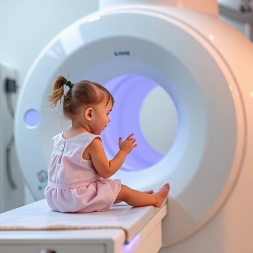
A groundbreaking study led by researchers from the University of California, San Francisco (UCSF) and UC Davis has unveiled a significant link between radiation exposure from medical imaging and increased risks of hematologic cancers in children and adolescents. This extensive investigation examined the health records of nearly four million children born between 1996 and 2016 across Canada and the United States, highlighting a concerning pattern of cancer risk associated with cumulative ionizing radiation doses attained via diagnostic imaging, particularly computed tomography (CT) scans.
The researchers meticulously analyzed vast datasets derived from six major healthcare systems in North America, providing a comprehensive retrospective cohort study that spanned two decades. Their findings identified that approximately one in ten blood cancers—equating to roughly 3,000 cases—may be attributable to the radiation children received during medically indicated imaging procedures. These results marked a pivotal advancement in pediatric oncologic epidemiology, substantiating a dose-dependent relationship where increased exposure translated to a heightened incidence of malignancies such as leukemia and lymphoma.
Central to the study is the distinction between different imaging modalities and their respective radiation doses. CT scans, especially head and brain CTs, were underscored for their relatively high levels of ionizing radiation, often necessary for acute diagnosis of conditions ranging from traumatic injuries to complex congenital anomalies. Contrastingly, conventional radiographs, such as chest X-rays, presented substantially lower radiation doses. The data revealed that children undergoing head CT scans faced nearly double the risk of hematologic cancers for one or two scans, surging to a 3.5-fold increased risk with multiple exposures, an alarming trend given the diagnostic ubiquity of these procedures.
Ionizing radiation, which includes X-rays used in medical imaging, interacts with cellular DNA and has long been established as a carcinogen. In pediatric populations, the vulnerability is accentuated due to the higher radiosensitivity of developing tissues and the longer lifespan available for potential cancer manifestations. The study affirms that this biological susceptibility necessitates heightened vigilance when applying imaging diagnostics, urging clinical protocols to strictly adhere to the principle of “as low as reasonably achievable” (ALARA) in radiation dosing. This concept champions tailoring scans to minimize exposure without compromising diagnostic detail.
Beyond statistical correlations, the authors delve into the mechanistic understanding of radiation-induced carcinogenesis. Ionizing radiation induces DNA double-strand breaks, chromosomal aberrations, and genomic instability, processes that can initiate malignant transformation in hematopoietic stem cells within the bone marrow. Given the high turnover rates and proliferative capacity of these progenitor cells in children, even low-dose exposures accumulate to potentially initiate oncogenesis. This mechanistic insight grounds the epidemiological findings in molecular biology, reinforcing the clinical implications.
Importantly, the study also advocates for the optimization of imaging strategies. It highlights the potential for substituting certain diagnostic procedures with non-ionizing modalities such as ultrasound and magnetic resonance imaging (MRI), which do not impart radiation and thus carry no associated carcinogenic risk. Such approaches, while sometimes limited by availability or diagnostic specificity, offer a pathway to reduce cumulative radiation exposures and mitigate the associated cancer risks.
In terms of demographic details, the study found that nearly 79% of the diagnosed hematologic cancers were lymphoid malignancies, while myeloid malignancies and acute leukemia accounted for approximately 15.5%. The data indicated a higher prevalence in males, with nearly half of all cases diagnosed before the age of five, underscoring the critical window of vulnerability during early childhood development.
The authors underline the indispensable role of medical imaging in pediatric medicine, recognizing its life-saving diagnostic and monitoring capabilities for myriad conditions. However, they stress the urgency of balancing these benefits with the long-term risks, particularly in an era where advanced imaging technologies have become increasingly accessible and frequently employed. This equilibrium requires multidisciplinary collaboration amongst radiologists, pediatricians, and health policy experts to set guidelines that prioritize patient safety without sacrificing clinical efficacy.
Dr. Rebecca Smith-Bindman, a leading radiologist and professor at UCSF, emphasizes that minimizing radiation dose during pediatric imaging is essential to safeguard children’s long-term health. She notes that children’s heightened radiosensitivity combined with their longer lifespan amplifies their risk for radiation-induced cancers, necessitating judicious use of CT scans and implementation of dose-reduction technologies. Additionally, Dr. Diana Miglioretti from UC Davis highlights the robust observed dose-response relationship, echoing international findings, and calls for clinicians to diligently weigh immediate diagnostic benefits against potential future harms.
The study’s funding sources included the National Cancer Institute, part of the National Institutes of Health, as well as support from Ontario’s Ministry of Health and Long-Term Care through the ICES health institute. Collaborators extended across prestigious institutions in the United States, Canada, and Australia, reflecting a broad and international commitment to understanding radiation risks in vulnerable pediatric populations.
These findings represent a crucial step toward refining pediatric imaging practices worldwide. The enhanced awareness cultivated by this research equips healthcare providers with empirical evidence to rigorously evaluate the necessity of radiation-based imaging and to pursue alternative diagnostic methods wherever feasible. It also fuels ongoing scientific inquiry into advanced dose reduction techniques and improved imaging protocols tailored for children.
Ultimately, this landmark study charts a path for safer pediatric healthcare by illuminating a significant public health concern linked to decades-old diagnostic practices. As clinicians integrate these insights, families and healthcare systems alike will benefit from improved guidelines that harmonize the imperatives of accurate diagnosis with the imperative to protect fragile developing tissues from preventable harm.
Subject of Research: Radiation exposure from medical imaging and its association with blood cancers in children
Article Title: Radiation from Pediatric Medical Imaging Linked to Increased Hematologic Cancer Risk: A Large-Scale Cohort Study
News Publication Date: September 17
Web References: UCSF News Release
References: The New England Journal of Medicine, National Cancer Institute grants R01CA185687 and R50CA211115, ICES Ontario Ministry of Health and Long-Term Care
Keywords: Medical imaging, Computed tomography (CT), Radiation, Pediatric hematology, Blood cancer, Leukemia, Lymphoma, Radiation-induced carcinogenesis, Pediatrics, Epidemiology, Radiation dose optimization, Ultrasound, MRI


