Scientists at Stanford University have made a groundbreaking advancement in the field of neuroscience with a novel method that allows researchers to view the intricate neural pathways in the developing brains of juvenile mice. This revolutionary approach involves a non-invasive technique that utilizes a compound, ampyrone, to render the scalp of the mouse transparent to light. The implications of this technique extend far beyond mere curiosity; it opens the door to exploring complex neurodevelopmental processes and forms of intervention for disorders that affect brain development.
Historically, studying the developing brain of juvenile mice has posed significant challenges for scientists. Traditional methods often involve sacrificing subjects for tissue analysis or using imaging techniques that are limited in scope and detail. The inability to repeatedly observe and record the same neural pathways over the span of weeks or months has stifled progress in understanding how these networks evolve. However, with the new technique, researchers can offset these limitations by simply applying ampyrone to the mouse’s scalp.
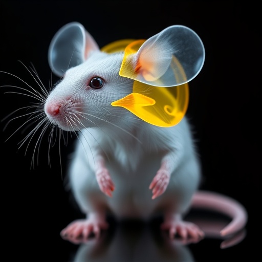
The transparency achieved through the application of ampyrone allows researchers to visualize the structures and activities of neurons in the juvenile mouse brain in remarkable detail. This achievement essentially creates a “window” into the brain. Guosong Hong, assistant professor of materials science and engineering and the senior author of the study, emphasized the transformative nature of this method, suggesting that it could pave the way for extensive research into the formation of neural circuits throughout development. Observing these processes in real time could prove essential for understanding how neural networks establish themselves and function effectively.
The mechanism behind this dramatic increase in visibility is rooted in the physics of light scattering. When light interacts with materials of varying optical properties, it scatters widely, limiting the clarity of images captured beneath the surface. In soft biological tissues, this scattering is prevalent, creating obstacles for researchers attempting to peer deeper into living organisms. The researchers identified that by adjusting the refractive index of water used in the solution, comparable to that of surrounding biological tissues, they could mitigate this scattering effect.
The concept of matching optical properties for enhanced visibility in biological systems is not new, but its successful application in live mice offers fresh promise. By incorporating ampyrone into the water and rubbing this solution onto the mouse’s scalp, researchers raised the refractive index sufficiently to achieve transparency, enabling them to capture data across the entire visible spectrum. This is particularly advantageous because the emitted fluorescence from genetically encoded markers in the neurons, such as yellow fluorescent protein (YFP), is essential for monitoring neuronal activity.
The findings related to neural imaging herald a significant step forward in the study of neurodevelopmental disorders, which remain poorly understood. With the ability to observe changes in neural circuits in live subjects over time, researchers can begin to understand the critical periods in brain development when plasticity is most pronounced. This knowledge could guide interventions for various conditions, including autism spectrum disorders and schizophrenia, where neurodevelopmental anomalies are often suspected to play a significant role.
Dr. Mark Brongersma, a co-author and professor of materials science and engineering, shared his fascination with the successful convergence of physics, chemistry, and biology. As a professional deeply engaged in the study of light-matter interactions, he remarked on the remarkable outcome of applying theoretical principles of optics to biological systems. The ability to “see” through previously opaque materials could fundamentally change how researchers explore biological phenomena.
The technique’s reversibility makes it particularly appealing for longitudinal studies. Unlike invasive techniques that can alter or damage the tissue, this method allows researchers to return to the same animal multiple times. This feature holds substantial promise for correlating developmental trajectories concerning environmental and genetic factors. The potential applications for understanding the basic principles of brain function and disorders are vast.
Past discoveries, such as the use of chemical agents to create transparency in red-light imaging, have set the stage for this latest advancement. The capability to transition from limited spectral visibility to the ability to see the full range of visible light dramatically enhances the versatility of imaging techniques employed in biomedical research. Researchers can now explore the details of various neural activities, potentially illuminating new pathways in cellular communication and signaling within the brain.
In practice, the researchers tested the method extensively, imaging both sedated and awake juvenile mice. Observing the neuronal activity, notably in response to sensory stimulation, provides insight into the fundamental workings of the mouse brain. The technique enables ongoing monitoring of changes over the course of development, and future studies may unlock additional understanding of how sensory experiences impact brain architecture.
The broad implications of this research extend into various scientific domains beyond neuroscience. Imagine how this technique could influence other areas requiring detailed imaging of soft tissues within live organisms. The potential for groundbreaking discoveries in the fields of pharmacology, regenerative medicine, and disease understanding cannot be understated.
In conclusion, the development of a method that allows researchers to non-invasively observe the brain’s neural pathways in juvenile mice represents a major leap forward in neuroscience research. With the newfound ability to visualize the formation and modification of neural circuits over time, scientists are poised to unravel complexities that could redefine our understanding of brain development and function. As they continue to refine this technique, there is excitement throughout the scientific community about the profound insights yet to be uncovered.
Subject of Research: Non-invasive imaging technique for observing neuronic pathways in juvenile mice
Article Title: Color-neutral and reversible tissue transparency enables longitudinal deep-tissue imaging in live mice
News Publication Date: 26-Aug-2025
Web References: Stanford News
References: Proceedings of the National Academy of Sciences
Image Credits: The Hong Lab
Keywords
Imaging, Neurodevelopmental Research, Optics in Biology, Neural Pathways, Mouse Models, Transparency Techniques, Light Scattering, Juvenile Mouse Brain, Fluorescent Imaging, Non-invasive Techniques, Neuroscience, Ampyrone.
