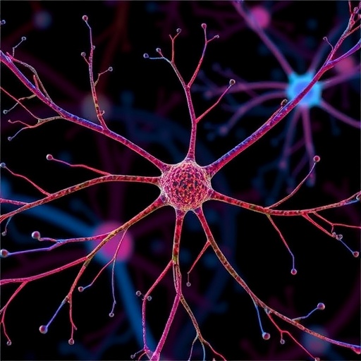
In the dynamic and complex environment of the cell, cytoskeletal structures such as microtubules play a critical role in shaping, supporting, and guiding cellular functions. These biopolymers, notable for their remarkable ability to self-organize, respond not only to biochemical signals but also to mechanical cues and spatial constraints within the cellular milieu. A recent groundbreaking study uncovers a novel mechanism by which microtubule networks adapt their growth and branching behavior in confined microenvironments, shedding light on processes that underpin neuronal development, plant morphogenesis, and fungal growth, with promising implications for bioengineering.
Microtubules (MTs) are filamentous polymers composed of tubulin subunits that constantly undergo phases of growth and shrinkage—a phenomenon known as dynamic instability. In living cells, MTs assemble into intricate networks that can be finely adjusted depending on the cellular context. Yet, the rules governing how these networks emerge, especially in geometrically constrained spaces such as narrow cellular protrusions, have remained elusive. Addressing this gap, a team led by Zaferani, Song, Wingreen, and colleagues systematically explored MT nucleation and branching within synthetic channels designed to mimic such tight cellular confines.
Their experimental platform utilized microfabricated channels featuring narrow junctions and closed ends to replicate the physical restrictions typical of axonal growth paths or dendritic extensions. Within these confined geometries, they observed that branching nucleation of MTs—a process whereby new filaments sprout from existing ones—is not uniform but highly sensitive to the spatial dimensions ahead of the microtubule’s growing tip. Specifically, the researchers discovered a previously uncharacterized form of mechanochemical feedback, which they termed “boundary sensing,” that governs whether branching initiation occurs beyond a bottleneck region.
This boundary sensing effect hinges upon a critical length threshold following narrow regions. When the distal space is sufficiently long, MTs extending toward the closed end undergo dynamic instability cycles, allowing a temporal window for new branching nucleation sites to arise further downstream. Conversely, when the distal compartment falls short of this minimum length, branching is suppressed, likely because the time for nucleation initiation does not outpace the depolymerization or catastrophe events at the MT tips near the boundary. This finding elegantly links spatial confinement with the biochemical kinetics of MT assembly, revealing a sophisticated intranetwork feedback.
Central to tuning this threshold is the branching factor TPX2, a cellular protein known to promote MT nucleation by recruiting γ-tubulin ring complexes that catalyze new filament formation. By modulating TPX2 levels within their system, the authors demonstrated that increasing the concentration accelerated branching rates and shortened the minimum length required for branching to emerge past narrow constrictions. However, when TPX2 was present in excess, an unexpected effect arose: MTs became highly stabilized at the closed end, which in turn obstructed the normal dynamic instability cycles and disrupted the formation of branched networks. This nuanced control highlights TPX2’s dual role as both an accelerator and a regulator of MT polymer dynamics in constrained microenvironments.
To complement their experimental observations, Zaferani and colleagues developed an integrative computational model simulating MT dynamics, nucleation kinetics, and boundary feedback mechanisms. Their simulations recapitulated the experimental trends, validating that the interplay between growth, catastrophe, and branching under confinement could be predicted quantitatively. These models provide powerful frameworks for predicting how cytoskeletal networks might adapt to complex, dynamic cellular geometries during processes such as axon elongation, dendritic arborization, or even specialized plant cell morphogenesis.
The broader implications of this work extend into developmental biology and applied bioengineering realms. In neurons, for instance, precise regulation of MT architecture within axonal and dendritic protrusions is essential for establishing functional connectivity and plasticity. The boundary-sensing mechanism revealed here offers a vital clue about how cells spatially organize their cytoskeleton to match morphological requirements dictated by physical constraints. Similarly, in plant and fungal cells exhibiting tip growth, MT network organization within confined tips is crucial for directional expansion and growth steering.
Moreover, this mechanistic understanding paves the way for designing biomaterials and synthetic tissues that harness cytoskeletal self-organization principles. By engineering channel geometries or tuning nucleation factors such as TPX2, it may become feasible to program the architecture of MT networks, thereby controlling cellular morphology and mechanical properties in tissue scaffolds or biohybrid devices. As the interface between biology and materials science continues to grow, insights into boundary sensing in MT networks will likely stimulate innovation in regenerative medicine and the creation of responsive biomimetic materials.
Another notable aspect of the study is its contribution to the fundamental understanding of cellular mechanosensation. Cells constantly perceive and respond to mechanical parameters such as tension, compression, and spatial confinement. Previous research had identified numerous biochemical pathways mediating mechanosensitive responses, but the direct coupling of MT polymerization dynamics with micron-scale geometrical constraints had not been elucidated in such quantitative detail. By directly linking the physical dimensions of confined spaces to nucleation kinetics, this work redefines microtubules as active agents capable of ‘sensing’ their spatial boundaries and adjusting their growth patterns accordingly.
Additionally, the role of dynamic instability emerges as a central player in enabling boundary sensing. The cycles of MT polymerization and depolymerization generate temporal fluctuations at filament ends, effectively providing a stochastic probe of the environment. Through these fluctuations, MTs can interpret the space available for growth, ensuring that branching nucleation only proceeds where it is spatially feasible. This insight reframes dynamic instability not merely as a source of cytoskeletal plasticity but as a key functional mechanism for spatial self-organization.
The research also emphasizes the importance of fine balance in cellular regulatory components. TPX2’s concentration must be carefully modulated to achieve desired branching and network morphology. This dependence mirrors cellular contexts where protein levels are tightly regulated via post-translational modification, degradation, or localized synthesis. Disruption of such balance may underlie pathological states where cytoskeletal organization is compromised, such as certain neurodegenerative diseases or cancer metastasis, pointing to the clinical relevance of these findings.
Importantly, the study leveraged a combination of cutting-edge in vitro reconstitution, microfabrication technologies, and advanced computational techniques—an interdisciplinary approach emblematic of modern cell biology. By bridging controlled experimental conditions with realistic geometries mimicking cellular compartments, and validating results with simulations, the authors set a new standard in dissecting cytoskeletal dynamics within physiologically relevant constraints. This methodology itself heralds a paradigm shift in exploring how biological polymers negotiate complex cellular landscapes.
Looking forward, several intriguing questions arise from this discovery. How do other cytoskeletal components, like actin filaments or intermediate filaments, integrate with this boundary-sensing mechanism? Can similar principles apply to multi-filament networks that collectively sustain cellular shape and motility? Furthermore, how do intracellular signaling pathways modulate TPX2 levels dynamically to orchestrate MT architecture during development or stress responses? These avenues promise fertile ground for future investigation stimulated by the present work.
In the realm of bioengineering, integrating boundary sensing principles into the design of artificial cellular systems or programmable materials could revolutionize how we harness cytoskeletal polymers for functional applications. For instance, biomimetic devices capable of adaptive remodeling upon encountering physical barriers could benefit from regulated branching nucleation systems inspired by microtubules. Such innovations may propel next-generation soft robotics, tissue engineering scaffolds, and environmental sensors.
In conclusion, the discovery of a boundary-sensing mechanism in branched microtubule networks that links spatial confinement with nucleation kinetics marks a significant advance in understanding cytoskeletal self-organization. By revealing how microtubules ‘read’ and adapt to their physical environment through dynamic instability and regulated branching, this research uncovers fundamental principles governing cellular architecture and morphogenesis. Coupled with insights into TPX2’s regulatory role and sophisticated modeling approaches, these findings open new frontiers across cell biology and bioengineering, promising impactful translational breakthroughs in health and technology.
Subject of Research: Self-organization and mechanosensing of branched microtubule networks under spatial confinement.
Article Title: Boundary-sensing mechanism in branched microtubule networks.
Article References:
Zaferani, M., Song, R., Wingreen, N.S. et al. Boundary-sensing mechanism in branched microtubule networks. Nat Chem Eng (2025). https://doi.org/10.1038/s44286-025-00264-0
Image Credits: AI Generated


