In a groundbreaking advancement with far-reaching implications for forensic science, a recent study has unveiled novel methodologies that leverage orthodontic profiles to enhance predictions of facial soft tissue thickness. This scientific breakthrough promises to refine the accuracy of forensic facial approximation—a crucial technique used in criminal investigations and identification procedures worldwide. By integrating detailed orthodontic data, researchers have charted a path toward reconstructing facial features with unprecedented precision, offering new hope in solving cold cases and identifying unknown individuals.
Forensic facial approximation—commonly known as forensic facial reconstruction—typically involves estimating the shape and volume of a person’s soft tissues, such as muscles and skin, based on skeletal remains. Traditionally reliant on population averages and limited anatomical markers, this method is inherently prone to uncertainties. However, the research spearheaded by Hanafi, Utsuno, Namiki, and colleagues, published in the International Journal of Legal Medicine, introduces orthodontic profiles as a potent predictive variable that can significantly narrow the margin of error in these reconstructions.
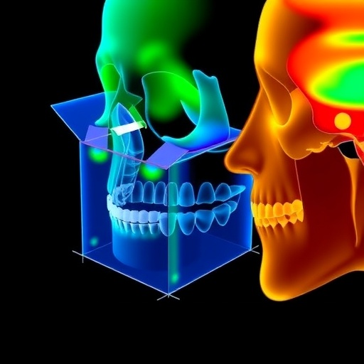
The study hinges on the fundamental understanding that the underlying dental and bone structures, particularly in the maxillofacial region, exert a direct influence on soft tissue contours. Orthodontic profiles, which detail the spatial orientation and alignment of teeth and jaws, provide a blueprint that reveals how soft tissues drape over the hard tissues beneath. This approach moves beyond conventional craniometric parameters, opening a new dimension for nuanced facial approximation.
The researchers meticulously compiled a dataset integrating cephalometric analyses—radiographic assessments that evaluate the head’s skeletal relationships—with 3D facial soft tissue measurements. Using advanced imaging technologies and statistical modeling, the team mapped correlations between distinct orthodontic variables and localized soft tissue thickness across multiple facial landmarks. This comprehensive mapping offers a layered understanding of how different dental arrangements correspond to specific soft tissue presentations.
One of the pivotal challenges addressed in the study is the variability in soft tissue thickness across diverse populations and age groups, which has historically complicated accurate reconstruction. By incorporating orthodontic variables, the team developed predictive models that account for this variability with greater granularity. Their models demonstrated statistically significant improvements in estimating tissue depths compared to standard anthropometric databases.
The implications of this are profound. Forensic artists and anthropologists can now utilize orthodontic profiles to generate facial approximations that are tailored not only to the skeletal traits but also to the subtle dental distinctions of an individual. This dual-layered approach improves the fidelity of facial reconstructions, making postmortem identifications more reliable and informative.
Furthermore, the research leverages cutting-edge computational tools and machine learning algorithms to analyze the multivariate data collected. By training algorithms on the orthodontic-soft tissue correlation matrices, the study introduces semi-automated predictive systems that can expedite and standardize forensic approximations. Such technological integration is a powerful step forward in modernizing forensic workflows.
Moreover, the interdisciplinary nature of this research exemplifies the synergy between orthodontics, forensic medicine, and computational science. It underscores the necessity of cross-field collaborations in tackling complex problems like accurate facial reconstruction. The orthodontic insights enrich forensic methodologies, while the forensic applications drive new orthodontic diagnostic perspectives.
Another key contribution of Hanafi and colleagues is their emphasis on validating their model across different ethnic backgrounds and age brackets. By doing so, they address the critical concern of applicability and universal relevance in forensic contexts that span global populations. This inclusivity ensures that the methodology does not fall short due to demographic biases—a common critique in anatomical research.
The authors also provide detailed technical discussions about the anatomical landmarks used and the precision of soft tissue thickness measurements. Coupled with high-resolution 3D scanning technology, they establish a rigorous framework for data acquisition that can be replicated and standardized globally. This methodological transparency enhances the study’s utility as a reference point for future research and forensic practice.
From a practical standpoint, law enforcement agencies, forensic laboratories, and medico-legal institutions stand to benefit immensely. The refined reconstructions derived from these enhanced models can improve public engagement and recognition, maximizing the chances of identifying missing persons or victims of crime. It is a tool that bridges the gap between skeletal remains and a recognizable human face.
It is noteworthy that the study also delves into potential future refinements, such as incorporating genetic data and further nuanced orthodontic variables. Such expansions hold the promise of tailoring approximations to an individual’s biogeographical ancestry or age-related morphologic changes, ushering in a new era of personalized forensic reconstruction.
Critics might argue that the integration of orthodontic data introduces complexity and necessitates specialized expertise. However, the standardization and automation efforts demonstrated mitigate this concern, making the technique accessible to forensic professionals with varied levels of orthodontic training.
The article also cautiously discusses limitations, including the necessity for high-quality radiographic data and comprehensive databases that represent the broad spectrum of human diversity. Addressing these limitations is part of the ongoing scientific evolution that this study catalyzes.
In conclusion, the incorporation of orthodontic profiles into forensic facial approximation represents a significant paradigm shift. The study by Hanafi and colleagues paves the way for more individualized, precise facial reconstructions that can profoundly impact forensic investigations and human identification globally. This fusion of anatomical detail with computational sophistication heralds an exciting frontier in forensic science.
As forensic science continues to evolve, the integration of diverse biological data sources with innovative analytical tools exemplifies the powerful trajectory toward ever-more accurate human identification techniques. The synergy between dental morphology and soft tissue modeling as explored in this research stands not only as a milestone in the field but as a beacon guiding the future of forensic facial reconstruction.
Subject of Research: Predicting facial soft tissue thickness for forensic facial approximation using orthodontic profiles.
Article Title: Assessing the contribution of orthodontic profiles in predicting facial soft tissue thickness for forensic facial approximation.
Article References:
Hanafi, M.G.S., Utsuno, H., Namiki, S. et al. Assessing the contribution of orthodontic profiles in predicting facial soft tissue thickness for forensic facial approximation. Int J Legal Med (2025). https://doi.org/10.1007/s00414-025-03542-x
Image Credits: AI Generated
