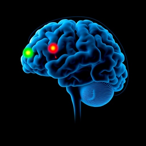
In a groundbreaking study published in npj Parkinson’s Disease, researchers have unveiled intricate details about the structural and functional transformations occurring in the posterior cerebellar vermis across various stages of Parkinson’s disease (PD), particularly focusing on gait dysfunction. This research sheds light on the cerebellum’s pivotal, yet often overlooked, role in the manifestation and progression of motor symptoms in PD, opening new avenues for targeted therapeutics aimed at alleviating one of the most debilitating aspects of the disease.
Parkinson’s disease, primarily characterized by its hallmark motor symptoms such as tremors, rigidity, and bradykinesia, also profoundly affects gait coordination, resulting in freezing of gait (FOG) and balance impairments that drastically reduce patient quality of life. Traditionally, research attention has concentrated on dopaminergic neuronal loss in the substantia nigra, but emerging evidence points to the cerebellum, particularly the posterior vermis, as a critical player in maintaining motor control integrity. The current study employs advanced neuroimaging and functional assessments to systematically map the extent and nature of regional-specific cerebellar alterations throughout PD progression.
The investigation utilized a combination of structural magnetic resonance imaging (MRI) and resting-state functional MRI (rs-fMRI) on cohorts representing early, middle, and advanced stages of PD exhibiting varying degrees of gait impairment. By dissecting volumetric changes alongside intrinsic connectivity patterns, the authors were able to delineate a complex picture wherein the posterior cerebellar vermis undergoes both morphological atrophy and functional reorganization paralleling the severity of gait dysfunction. These findings challenge the traditional notion of cerebellar preservation in PD and highlight a dynamic and regionally selective cerebellar involvement.
Morphometric analyses disclosed a pronounced reduction in gray matter volume specifically localized within the lobules VI and VII of the posterior vermis. These lobules have been implicated in sensorimotor processing and postural control, which are essential for coordinated locomotion. Notably, the degree of atrophy was significantly correlated with clinical assessments of gait impairment and freezing episodes, suggesting a direct link between structural degeneration and motor symptomatology. This revelation enhances our understanding of cerebellar vulnerability in PD and underlines the anatomical specificity underlying gait disturbances.
Beyond structural decline, resting-state functional connectivity analyses unveiled profound disruptions within intrinsic cerebellar networks as well as altered cerebellar-cortical communication pathways. Particularly, diminished connectivity between the posterior vermis and primary motor cortex, supplementary motor area, and basal ganglia circuits was observed. These functional decouplings were more severe in advanced stages and correlated with worsening gait metrics. Such findings suggest that the cerebellum’s integrative role in sensorimotor feedback loops is compromised, potentially precipitating the characteristic locomotor deficits observed in PD.
The study further elucidated compensatory mechanisms engaged in early disease stages, where hyperconnectivity within certain cerebellar subregions possibly represents an adaptive response to initial dopaminergic deficits. However, as neurodegeneration progresses, these compensatory networks fail, leading to network breakdown and manifest clinical impairments. This biphasic functional trajectory underscores the plasticity of cerebellar networks and highlights critical windows for therapeutic interventions aimed at bolstering cerebellar resilience or modulating aberrant connectivity.
A critical methodological strength of the study lies in its longitudinal design, capturing cerebellar alterations across disease time course rather than at single time points. This temporal perspective is vital in understanding PD’s evolving neuropathology, as it reveals not only static changes but also dynamic remodeling processes that may inform staging and prognosis. Such an approach is poised to advance biomarker development, offering refined neuroanatomical signatures of gait dysfunction progression.
This research carries profound clinical implications. Understanding the cerebellar vermis involvement provides a neurobiological substrate for developing novel neuromodulation therapies, such as targeted transcranial magnetic stimulation (TMS) or deep brain stimulation (DBS) protocols, tailored to normalize cerebellar-cortical network dysfunction. Moreover, it invites reconsideration of rehabilitative strategies focusing on sensorimotor integration, balance training, and neuroplasticity enhancement to ameliorate gait deficits through cerebellar engagement.
Importantly, the findings challenge the dopamine-centric therapeutic paradigm that dominates PD management. By highlighting the cerebellar contribution to motor symptoms, the study advocates for a more holistic view encompassing multi-system neurodegeneration and multisite functional disturbances. Such an integrated framework is critical for addressing complex symptoms like freezing of gait, which are refractory to conventional dopaminergic treatments.
The authors acknowledge limitations, including variability in medication regimens potentially influencing functional connectivity patterns and the cross-sectional nature of some data points. Future investigations incorporating larger samples and multimodal imaging, including diffusion tensor imaging for white matter integrity assessment, could further elucidate the cerebellar circuitry changes. Additionally, integrating clinical gait analysis with neuroimaging may refine our understanding of cerebellar contributions to specific motor subphenotypes.
As the field moves forward, this study serves as a clarion call for revisiting cerebellar involvement in neurodegenerative frameworks beyond PD, such as atypical parkinsonian syndromes and other movement disorders. It pioneers a model wherein region-specific cerebellar alterations inform both pathophysiological understanding and personalized medicine approaches.
In conclusion, the meticulous examination of the posterior cerebellar vermis presents a paradigm shift in conceptualizing Parkinson’s disease as a multisystem disorder with significant cerebellar pathology underlying gait dysfunction. This research paves the way for novel diagnostic markers and targeted therapeutics, offering hope for improving mobility and quality of life in patients burdened by the relentless progression of Parkinson’s disease.
Subject of Research: Parkinson’s disease progression and gait dysfunction related to structural and functional changes in the posterior cerebellar vermis.
Article Title: Regional-specific structural and functional changes of posterior cerebellar vermis across different stages of Parkinson’s disease with gait dysfunction.
Article References:
Yu, L., Han, J., Chen, X. et al. Regional-specific structural and functional changes of posterior cerebellar vermis across different stages of Parkinson’s disease with gait dysfunction. npj Parkinsons Dis. 11, 208 (2025). https://doi.org/10.1038/s41531-025-01065-1
Image Credits: AI Generated


