Forensic anthropologists evaluating skeletal remains from a Northern Thai population have demonstrated that morphometric dimensions of the tibia, particularly measurements taken from the proximal segment and the tibial tuberosity, provide highly accurate mathematical models for osteometric sex estimation. Investigating two hundred and forty skeletal elements curated at Chiang Mai University, researchers determined that multivariate discriminant function equations incorporating complete tibial variables reached ninety-nine point five percent classification accuracy in initial modeling and one hundred percent accuracy within an independent holdout sample. The investigation specifically established that the tibial tuberosity, an anterior bony projection influenced by patellar tensile forces, functions as a reliable salvage indicator when trauma, taphonomic degradation, or mass disasters compromise more fragile skeletal structures.
The identification of biological sex represents an indispensable foundational phase in the reconstruction of an unknown decedent’s biological profile during medicolegal investigations. While the adult human pelvis consistently offers the highest diagnostic reliability across skeletal collections, followed closely by cranial landmarks, these anatomical regions are notoriously susceptible to severe postmortem fragmentation, crushing, and accelerated environmental decay. Consequently, forensic researchers have increasingly directed attention toward the postcranial appendicular skeleton, where large long bones often demonstrate marked sexual dimorphism and preserve structural integrity across extended postmortem intervals due to their substantial cortical thickness and biomechanical density.
As a primary weight-bearing pillar transmitting biomechanical loads between the knee and ankle joints, its epiphyses and diaphyseal regions develop prominent morphological landmarks shaped by continuous locomotor stress, body weight, and muscular traction.
To establish rigorous, population-specific forensic standards for Northern Thailand, the research team analyzed two hundred and forty adult left tibiae curated at the Osteology Research and Training Center within the Faculty of Medicine at Chiang Mai University. The skeletal sample comprised equal numbers of male and female individuals, with one hundred and twenty specimens per sex. Prior to primary modeling, the investigators conducted a preliminary bilateral investigation utilizing forty paired left and right tibiae evaluated through paired-sample t-tests, which revealed no statistically significant bilateral asymmetry between sides, thereby justifying the exclusive analytical focus on the left tibia. The experimental cohort was structured into a model development training set consisting of two hundred left tibiae, comprising one hundred males and one hundred females, and a separate holdout validation cohort of forty specimens, containing twenty males and twenty females. The documented age at death across the entire sample spanned from twenty-six to ninety-four years, exhibiting mean ages of fifty-three point four zero years for males and fifty-four point six zero years for females, while all fractured, incomplete, or severely pathologically altered bones were systematically excluded.
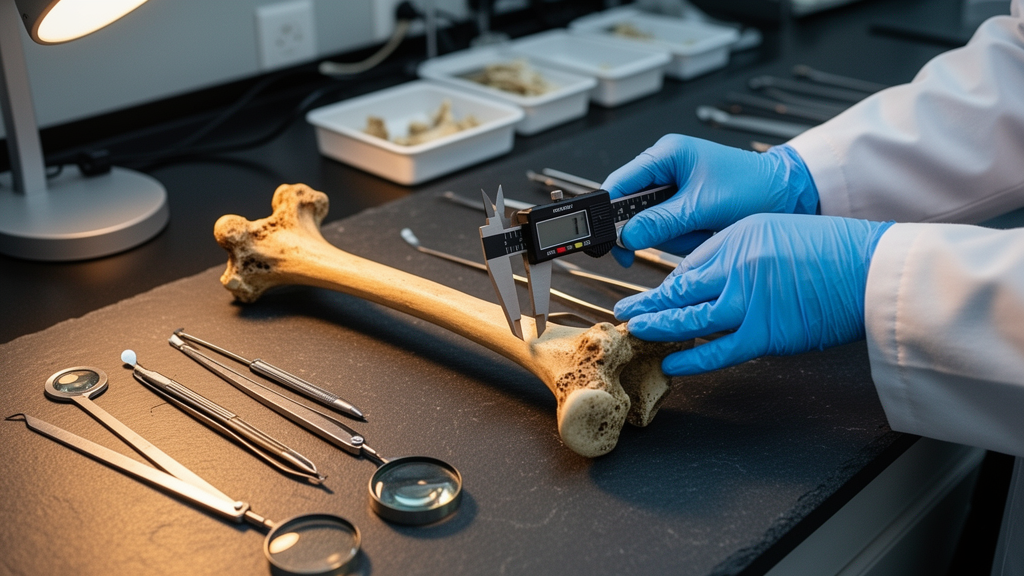
The experimental protocol encompassed nine distinct morphometric dimensions recorded to the nearest zero point zero one millimeter using digital sliding calipers, a specialized osteometric board, and flexible osteometric tape. Six conventional tibial metrics were gathered alongside three targeted dimensions of the tibial tuberosity. The standardized parameters included total tibial length, upper articular breadth measured across the proximal epicondylar articular rims, and lower articular breadth taken across the distal articular surface from the fibular notch to the medial malleolus. Diaphyseal dimensions comprised the anteroposterior diameter, transverse diameter, and maximum shaft circumference, all recorded precisely at the level of the nutrient foramen. To quantify the tibial tuberosity as a discrete forensic indicator, researchers recorded its longitudinal diameter from the proximal bulging margin to the distal boundary, its transverse diameter at mid-length, and the anterior height measured from the mid-proximal articular margin to the most projecting anterior point of the tuberosity.
To guarantee strict measurement precision and eliminate intra-examiner drift, all primary osteometric assessments were recorded in triplicate by the initial investigator, with arithmetic means utilized for statistical modeling. Measurement reliability and reproducibility were formally evaluated through the technical error of measurement, relative technical error of measurement, and the coefficient of reliability across forty randomly selected specimens remeasured after a four-week interval. Independent evaluation was simultaneously executed by a secondary qualified forensic anthropologist who remained fully blinded to original measurements to establish true inter-observer reproducibility. Following normality verification via the Kolmogorov-Smirnov test, the authors applied independent-sample t-tests to evaluate sexual dimorphism, followed by direct and stepwise discriminant function analyses in SPSS version twenty-six to construct predictive univariate and multivariate mathematical equations capable of classifying unknown fragmentary bones.
Statistical evaluation confirmed that every measured tibial parameter adhered to normal distribution curves and displayed highly significant sexual dimorphism, with male specimens demonstrating statistically significantly larger dimensions across all nine parameters at probability values below zero point zero one. In multivariate discriminant function modeling, the complete-tibia equation incorporating all measured variables achieved an initial classification accuracy of ninety-nine point five percent. A regional proximal model utilizing four proximal parameters maintained a ninety-eight point zero percent classification accuracy under both original testing and leave-one-out cross-validation routines. An equation based solely on the three tibial tuberosity variables also attained an impressive ninety-eight point zero percent accuracy, whereas the diaphyseal shaft model produced an acceptable but comparatively reduced original accuracy of eighty-one point five percent.
When these derived mathematical functions were applied blindly to the forty independent holdout specimens, multivariate equations incorporating complete, proximal, and tibial tuberosity dimensions each demonstrated one hundred percent correct classification accuracy. The calculated ninety-five percent confidence intervals using the Wilson score method ranged from ninety-one point two percent to one hundred percent, confirming the strong diagnostic power of the proximal third of the bone. Among single-variable univariate models, upper articular breadth emerged as the single most effective individual discriminator, yielding a holdout accuracy of ninety-five point zero percent with a confidence interval spanning eighty-three point five to ninety-eight point six percent. Lower articular breadth and the longitudinal diameter of the tibial tuberosity each achieved ninety point zero percent classification accuracy on the holdout specimens, while the shaft circumference at the nutrient foramen attained eighty-seven point five percent.
The technical evaluation of measurement error revealed meaningful contrasts between well-defined articular landmarks and the less distinct boundaries of the anterior tuberosity. Intra-observer reliability was consistently high across all variables, with reliability coefficients exceeding zero point nine one, and most exceeding zero point nine five. Inter-observer reproducibility for standardized skeletal landmarks, including total length, proximal articular breadth, distal breadth, and all nutrient foramen shaft dimensions, proved exceptional, demonstrating reliability coefficients between zero point nine five and one point zero zero alongside relative error rates below three percent. Conversely, dimensions involving the margins of the tibial tuberosity displayed greater inter-examiner discordance. The longitudinal diameter of the tuberosity exhibited the lowest inter-observer coefficient of reliability at zero point seven six with a relative technical error of six point four five percent, while transverse diameter and anterior height produced reliability coefficients of zero point seven nine and zero point eight seven, respectively.
These variations in inter-observer agreement stem directly from the underlying morphological nature of the tibial tuberosity as an anatomical traction apophysis rather than a clearly demarcated articular facet. While joint surfaces like the proximal condyles possess discrete osteometric margins that facilitate precise caliper placement, the peripheral borders of the tuberosity blend gradually into the anterior crest of the tibial diaphysis without an abrupt border. Furthermore, as an attachment site subjected to repetitive tensile loading from the quadriceps muscle group and patellar ligament during locomotion, the tuberosity undergoes dynamic bone remodeling influenced by individual biomechanical stress, body mass index, and physical activity. Consequently, an athletic female may develop a more pronounced tuberosity than a sedentary male, introducing biological variation that can potentially confound purely metric sex allocation if evaluated in complete isolation.
Because the tibial tuberosity is reinforced by dense, thick underlying cortical bone, it frequently survives taphonomic crushing and carnivore scavenging that preferentially destroy marrow-rich cancellous epiphyses. In forensic practice, the authors therefore advocate for a hierarchical diagnostic framework where the tuberosity is treated primarily as a secondary salvage variable or integrated into multivariate discriminant functions rather than functioning as an unassisted primary classifier. When pristine articular margins like the upper articular breadth are preserved, they should remain the primary metric reference points; however, when taphonomic forces leave only fragmentary anterior fragments intact, the validated tuberosity metrics provide dependable identification evidence that substantially outperforms chance and overcomes minor technical measurement inconsistencies.
The investigators emphasized important methodological caveats, noting that the perfect one hundred percent accuracy observed across several holdout models reflects an expected statistical anomaly related to the constrained size of the forty-specimen validation sample rather than absolute biological certainty across the entire Thai population. The dynamic biomechanical nature of the tuberosity also introduces morphological traits, such as enthesophytes or changes associated with adolescent Osgood-Schlatter disease, which may alter dimensions while simultaneously offering distinct markers for individualization against antemortem medical records. To overcome manual caliper subjectivity and further refine population databases, future research directions include the application of computed tomography, three-dimensional digital imaging, and deep learning algorithms to capture living anatomical variations, thereby expanding the forensic toolkit for analyzing fragmentary human remains.
Subject of Research: Forensic osteometric sex estimation from tibial morphometry in a Thai population
Article Title: Sex estimation from the tibia in Thais: validation of tibial tuberosity as a novel osteometric indicator
Article References: Malawan, P., & Mahakkanukrauh, P. (2026). Sex estimation from the tibia in Thais: validation of tibial tuberosity as a novel osteometric indicator. Egyptian Journal of Forensic Sciences, 16(1), Article 65. https://doi.org/10.1186/s41935-026-00565-2
Image Credits: AI Generated
DOI: 10.1186/s41935-026-00565-2
Keywords: forensic anthropology, sex estimation, tibial morphometry, tibial tuberosity, osteometrics, discriminant function analysis, skeletal identification, Thai population, forensic osteology, upper articular breadth, taphonomy, fragmentary remains, technical error of measurement, biological profile, Chiang Mai University, holdout validation
Cite Scienmag News
Peter Mason. (November 12, 2026). Thai Tibia Study Validates Tibial Tuberosity for Forensic Sex Estimation. Scienmag. https://scienmag.com/thai-tibia-study-validates-tibial-tuberosity-for-forensic-sex-estimation/
Peter Mason. "Thai Tibia Study Validates Tibial Tuberosity for Forensic Sex Estimation." Scienmag, 12 November 2026, https://scienmag.com/thai-tibia-study-validates-tibial-tuberosity-for-forensic-sex-estimation/. Accessed 12 November 2026.
Peter Mason. "Thai Tibia Study Validates Tibial Tuberosity for Forensic Sex Estimation." Scienmag. November 12, 2026. https://scienmag.com/thai-tibia-study-validates-tibial-tuberosity-for-forensic-sex-estimation/