


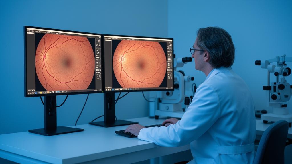
Under standard downstream evaluation settings, a computational framework termed Multi-View Self-Supervised Learning, or MVSSL, achieves an 82.72 ± 0.23 percent Dice score on STARE retinal vessel segmentation and an 85.03 ± 0.98 percent Dice score on RIM-ONE-r3 optic cup segmentation. These measured segmentation outcomes, reported in a study published in Applied Intelligence by researchers Shumao Wang, Ximeng Zhang, and Song Guo, demonstrate improved performance over the representative baseline VICRegL, which recorded 81.75 ± 0.45 percent Dice and 84.16 ± 0.53 percent Dice on those respective tasks. Across comprehensive testing, the authors report that the proposed MVSSL framework yields competitive and generally improved performance over representative self-supervised learning baselines for retinal fundus image segmentation tasks.
The investigation focuses on addressing key limitations in conventional self-supervised learning approaches applied to retinal fundus image segmentation. Self-supervised learning provides an effective mechanism to learn transferable representations from unlabeled retinal images, thereby reducing the dependence of downstream segmentation tasks on manual annotations. However, existing conventional self-supervised methods often rely predominantly on global image views. As the authors note, this reliance on global perspective can prove insufficient for retinal fundus images containing anatomical structures and pathological lesions that appear at substantially different spatial scales across the visual field.
To address this issue, the researchers developed the Multi-View Self-Supervised Learning framework to jointly learn representations from wide field-of-view and narrow field-of-view inputs. The architecture separates the input processing across two dedicated branches designed for distinct spatial resolutions. The wide field-of-view branch processes larger retinal regions to capture broader contextual patterns across the fundus. Meanwhile, the narrow field-of-view branch uses multiple local views together with feature-space correspondence matching. This specialized correspondence matching in the feature space is specifically implemented to facilitate fine-grained representation learning for subtle local structures.
In addition to the multi-view dual-branch structure, the framework introduces grid shuffling as an input-level structural perturbation. The primary objective of introducing grid shuffling is to reduce the model’s reliance on fixed spatial layouts while preserving the underlying local visual content of the images.
The authors evaluated the performance of the proposed MVSSL framework across five distinct downstream retinal segmentation tasks. These target applications encompassed the segmentation of retinal blood vessels, optic discs, optic cups, hard exudates, and microaneurysms.
Beyond standard evaluations, further experiments conducted under few-shot and cross-domain settings demonstrated favorable transferability for the learned representations. To assess compatibility with alternative segmentation architectures, the researchers carried out additional experiments using U-Net++, which demonstrated that the learned representations remain effective when paired with a more advanced segmentation decoder. The authors have made the underlying source code accessible through GitHub at https://github.com/guomugong/MVSSL.
Subject of Research: Development of a multi-view self-supervised learning framework for retinal fundus image segmentation
Article Title: Multi-view self-supervised learning with grid shuffling for retinal fundus image segmentation
Article References: Wang, S., Zhang, X., & Guo, S. (2026). Multi-view self-supervised learning with grid shuffling for retinal fundus image segmentation. Applied Intelligence, 56(16), Article 535. https://doi.org/10.1007/s10489-026-07571-w
Image Credits: AI Generated
DOI: 10.1007/s10489-026-07571-w
Keywords: self-supervised learning, retinal segmentation, medical imaging, fundus photography, computer vision, deep learning, grid shuffling, multi-view learning, optical coherence tomography, DICOM, Multi-view, self-supervised
Cite Scienmag News
Blake Davidson. (November 11, 2026). New Framework Improves Retinal Image Segmentation via Multi-View Learning. Scienmag. https://scienmag.com/new-framework-improves-retinal-image-segmentation-via-multi-view-learning/
Blake Davidson. "New Framework Improves Retinal Image Segmentation via Multi-View Learning." Scienmag, 11 November 2026, https://scienmag.com/new-framework-improves-retinal-image-segmentation-via-multi-view-learning/. Accessed 11 November 2026.
Blake Davidson. "New Framework Improves Retinal Image Segmentation via Multi-View Learning." Scienmag. November 11, 2026. https://scienmag.com/new-framework-improves-retinal-image-segmentation-via-multi-view-learning/




