


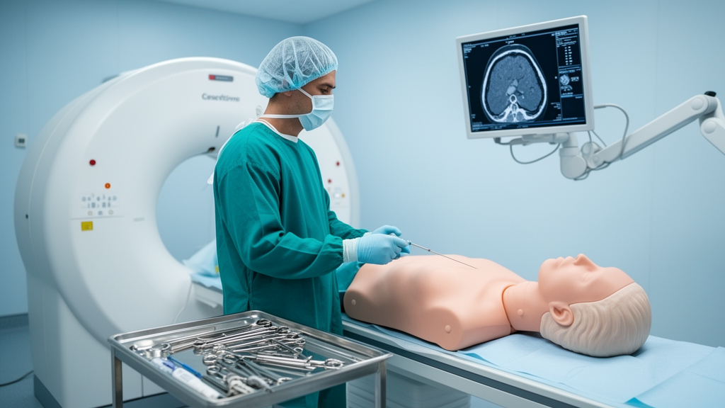
An automated image-processing tool designed to track procedural needles on computed tomography scans significantly accelerated targeting times and decreased the number of interim verification images required during simulated interventional procedures, according to a prospective phantom study published in CVIR Oncology. Conducting sixty needle placements across a standardized torso model, an investigative team evaluated the operational impact of specialized Needle Detection software compared directly against conventional freehand CT guidance. The controlled multi-operator trial revealed that real-time algorithmic tracking cut the average time required to reach internal targets by nearly half, while simultaneously reducing the frequency of intermediate control scans. Because the trial took place within an inanimate abdominal phantom rather than living clinical subjects, direct radiation exposure was not measured, but the systematic reduction in required imaging acquisitions highlights the operational potential of software-guided interventions.
Computed tomography-guided percutaneous needle placement serves as an indispensable tool across modern interventional radiology, forming the technical foundation for essential diagnostic biopsies, fluid collections drainage, local tumor ablations, and targeted pain therapy interventions. Interventional practitioners rely extensively on CT imaging due to its high spatial resolution, predictable anatomical delineation, and widespread availability in hospital environments. Advancing a fine needle toward small, deeply embedded, or obliquely oriented anatomical structures remains technically demanding, however, even for highly practiced clinicians. Under current clinical standards, physicians must repeatedly execute iterative cycles of manual needle advancement followed by interim axial imaging to confirm trajectory alignment, depth, and spatial orientation relative to delicate critical anatomy.
These stepwise manual adjustments introduce procedural interruptions and prolong intervention durations, which can directly affect patient tolerance and clinical suite throughput. Every additional interim verification scan acquired during needle progression adds to the patient’s cumulative radiation exposure, presenting a persistent concern in radiological workflows. Although low-dose spiral protocols and intermittent scanning techniques have substantially diminished the radiation burden per individual acquisition, overall exposure remains directly proportional to the total number of control scans performed. Complex access trajectories, such as steep out-of-plane angulations or long in-plane paths, exacerbate these difficulties. Furthermore, needle visibility is frequently impaired by physical factors including beam-hardening artifacts, metal-related scattering, partial volume averaging, and unfavorable soft-tissue contrast conditions, forcing operators to execute repeated repositioning maneuvers that further extend procedure times.
To mitigate the limitations of conventional guidance, medical device developers have introduced diverse technological platforms, including CT fluoroscopy, electromagnetic navigation systems, robotic assistance units, and augmented reality headsets. While such modalities can enhance navigational precision, they routinely demand costly auxiliary hardware, substantial procedural room reconfigurations, extensive operator retraining, or heightened occupational radiation exposure for interventional staff remaining tableside. By comparison, dedicated software-based needle guidance represents an integrated workflow alternative. These software platforms aim to improve visualization and spatial localization directly on standard CT images without necessitating specialized external instrumentation, hardware recalibration, or modifications to routine clinical imaging acquisition protocols.
The investigated software platform, identified as myNeedle Detection (version SYNGO CT VB20A), operates by deploying specialized image-processing algorithms to automatically identify, enhance, and continuously track high-density linear metallic structures corresponding to the needle shaft and tip. Throughout the intervention, the system continuously updates needle visualization following every interim scan and superimposes a graphic representation of the planned path established by the radiologist based on the initial entry point and target location. Beyond visual trajectory overlays, the interface computes and displays dynamic quantitative metrics in real time. These metrics provide interventionalists with the remaining distance to the target in millimeters alongside precise angular deviations across the lateral x-axis and vertical z-axis, allowing clinicians to make rapid, objective trajectory corrections between physical advancements.
To evaluate the tool under rigorous, reproducible conditions, the researchers utilized a standardized multi-modality abdominal phantom (model 057 A, CIRS, Norfolk, VA, USA) incorporating six predefined internal targets. These targets were engineered to represent varied tiers of procedural complexity, differing in intraphantom depth from superficial to deep locations and featuring both single-angled and challenging double-angled trajectories across two distinct spatial planes. These varied configurations required diverse degrees of needle manipulation and spatial orientation, simulating access challenges encountered in actual clinical settings. By utilizing an inanimate torso phantom with predefined skin entry markers and fixed internal coordinates, the study established balanced task difficulty while deliberately excluding patient-related confounders such as respiratory excursions, involuntary physical motion, tissue deformation, and active hemorrhage.
All puncture procedures were conducted using a multidetector CT scanner (SOMATOM X.Ceed, Siemens Healthineers) equipped with the myNeedle Guide 3D software application and an accompanying myNeedle Laser positioning system. Image acquisitions adhered to a standardized low-dose spiral intervention protocol set at a tube voltage of 125 kVp and an effective tube current of approximately 80 mAs. Under these standardized scanner settings, each verification scan yielded a volume CT dose index (CTDIvol) of approximately 2.4 mGy. Operators advanced needles in an incremental stepwise fashion, requesting interim verification spiral scans at their professional discretion to confirm path fidelity. The study placed no artificial restrictions on needle repositioning frequency, angular adjustments, or total scanning events, thereby mirroring standard interventional decision-making paradigms.
The study cohort comprised ten radiologists possessing a broad spectrum of technical expertise to assess whether guidance benefits were tied to operator experience. Three participants had six years of experience in CT-guided interventions, two had five years, two had two years, and three operators were novices with no prior hands-on puncture experience. Puncture sequences and software activation states were balanced and randomized for each operator to eliminate procedural order bias and mitigate progressive operator learning curves. Prior to performing the experimental insertions, all ten operators received a brief, standardized orientation covering the software’s interface features and quantitative coordinate readouts.
Across all sixty procedures, operators attained a technical success rate of 100%, with computed tomography confirming successful needle tip placement within the predefined target in every attempt without technical failures or data loss. Analysis of procedural time demonstrated marked efficiency gains under software assistance. The mean time to target decreased from 422.5 ± 199.8 seconds during standard guidance to 224.3 ± 103.3 seconds when utilizing the needle detection tool, representing an approximate 47% reduction in overall procedure duration. Nonparametric comparison via the Mann–Whitney U test confirmed that this decrease was statistically significant (p < 0.001). Median durations exhibited a parallel drop, declining from 336.5 seconds without software support to 209.5 seconds with active detection, reflecting an unambiguous distributional shift toward expedited targeting.
The frequency of interim imaging verification scans required during needle advancement was likewise significantly curtailed by the tracking software. Under conventional CT guidance, operators required a median of 4 verification scans (interquartile range: 3 to 6) to reach the intended target, whereas software-assisted guidance required a median of 3 verification scans (interquartile range: 2 to 4). Statistical evaluation using a permutation test with 10,000 iterations confirmed the significance of this reduction (p < 0.0001). The 10,000 permutations served strictly to calculate the null distribution of the test statistic rather than inflating sample observations. Sensitivity analyses incorporating linear mixed-effects modeling for procedure duration and generalized linear mixed Poisson modeling for scan counts, each using random intercepts for individual operators, reaffirmed the statistical robustness of the primary findings.
Crucially, the observed operational advantages manifested across all ten participating radiologists, regardless of prior interventional background. Although baseline execution metrics naturally varied among individual practitioners, every single radiologist registered shorter targeting times and fewer interim verification scans when utilizing the needle detection application. The authors noted that for highly experienced interventionalists, the automatic trajectory annotations and continuous angular readouts likely reduced cognitive burden and streamlined spatial adjustments.
Despite these encouraging findings, the study authors emphasize several vital methodological limitations that preclude immediate clinical generalization. Because experiments were conducted strictly within a solid synthetic phantom, the study could not capture real-world patient dynamics, including respiratory lung sliding, cardiac motion, tissue compliance changes, internal organ shifting, or procedural bleeding. Furthermore, the trial was designed specifically to examine workflow efficiency rather than millimeter-level targeting accuracy, leaving questions open regarding whether software tracking improves final tip precision. Procedural radiation was evaluated solely through the surrogate metric of scan frequency under identical acquisition parameters; direct radiation dosimetry was not performed. Consequently, the investigators caution that fewer scans must not be interpreted as definitive proof of decreased patient radiation dose until prospective clinical trials incorporating direct dosimetric measurements are conducted.
Subject of Research: Procedural efficiency and verification scan frequency of CT needle detection software in a phantom model
Article Title: Needle Detection software improves procedural efficiency and reduces verification scans in CT-guided needle placement: a multi-operator phantom study
Article References: Saeed, S., Niehoff, J. H., Roettcher, T., Schreck, J., Lueking, J., Schoenbeck, D., Katz, M. S., Mendorf, A., Ottemoeller, J., Muecke, R., Almatar, M., Shekho, D., Moenninghoff, C., Borggrefe, J., & Kroeger, J. R. (2026). Needle Detection software improves procedural efficiency and reduces verification scans in CT-guided needle placement: a multi-operator phantom study. CVIR Oncology, 2(1), Article 38. https://doi.org/10.1007/s44343-026-00071-1
Image Credits: AI Generated
DOI: 10.1007/s44343-026-00071-1
Keywords: interventional radiology, computed tomography, needle guidance, phantom study, image-processing algorithm, procedural efficiency, verification scans, radiation surrogate, needle detection software, Needle, Detection, software
Cite Scienmag News
Nathaniel Bowman. (November 9, 2026). Needle Detection Software Accelerates CT-Guided Interventions in Torso Phantom Study. Scienmag. https://scienmag.com/needle-detection-software-accelerates-ct-guided-interventions-in-torso-phantom-study/
Nathaniel Bowman. "Needle Detection Software Accelerates CT-Guided Interventions in Torso Phantom Study." Scienmag, 9 November 2026, https://scienmag.com/needle-detection-software-accelerates-ct-guided-interventions-in-torso-phantom-study/. Accessed 9 November 2026.
Nathaniel Bowman. "Needle Detection Software Accelerates CT-Guided Interventions in Torso Phantom Study." Scienmag. November 9, 2026. https://scienmag.com/needle-detection-software-accelerates-ct-guided-interventions-in-torso-phantom-study/




