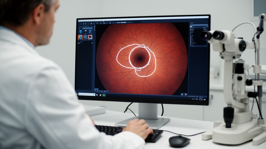
Glaucoma steals sight quietly. The disease damages the optic nerve gradually, and patients often notice nothing until the loss becomes irreversible, which is why early detection matters so much. A key step in that detection is measuring the cup-to-disc ratio, a number derived from how deeply the optic cup sits within the optic disc at the back of the eye. Computing this ratio reliably requires precisely outlining both structures in retinal photographs, a task that is notoriously difficult because the cup’s boundary is faint, low in contrast, and frequently crossed by blood vessels. A new study published in BMC Ophthalmology presents a deep learning framework, called EEGA-VMUNet, that tackles this segmentation problem and reports competitive accuracy across four public benchmark datasets.
The research team, led by Lili Cai, Keke Hou and Zhong Mo, built their model on a relatively recent architectural idea: the Mamba state space model. Unlike transformers, which rely on self-attention whose computational cost grows sharply with image size, Mamba-based designs model long-range dependencies with computational demands that scale linearly with input length. The authors adopted the Vision Mamba UNet, or VM-UNet, as their backbone. Its encoder and decoder are both equipped with a visual state space mechanism, in which a two-dimensional selective scan module sweeps images along horizontal, vertical and diagonal directions to capture global context efficiently. For optic disc and cup segmentation, this global view matters: when local intensity cues fail at an obscured cup boundary, information about the overall shape and size of the disc, and the relative spatial positions of disc and cup, can supply the missing context.
On top of this backbone, the researchers introduced their main novelty, an enhanced edge-guided attention module. Building on an existing edge-guided attention framework, they replaced part of the original structure with a global attention mechanism and added a dual attention module, creating two parallel hybrid attention branches whose outputs are summed. The module sits in the skip connections between encoder and decoder, fusing encoded features, decoded features and an edge feature map produced by Laplacian pyramid decomposition. According to the authors, this design preserves channel and spatial information, suppresses vascular noise, and sharpens the representation of the fine boundaries where segmentation errors concentrate. Ablation experiments suggested that both attention branches contribute: removing the global attention branch lowered the cup Dice score from 92.23 percent to 91.28 percent on one dataset, and removing the dual attention branch lowered it to 91.63 percent.
The framework also includes a practical preprocessing stage. Fundus photographs are high-resolution, and most of each image is background, so feeding full images into a segmentation network wastes computation and can hurt accuracy. The team used a YOLOv10 object detector, trained on 400 full-field images from the REFUGE dataset, to localize the optic disc and crop a region of interest, which is then resized to 256 by 256 pixels. Because the cup lies anatomically within the disc, localizing the disc captures both structures. On the REFUGE validation set, the detector reached a mean average precision at 0.5 intersection-over-union of 0.9944, with precision of 0.9924 and recall of 0.9788. A 30 percent padding margin around each detected box ensured the cropped region fully contained the optic disc in 100 percent of REFUGE images, absorbing minor localization errors.
Training relied on a multi-scale loss function combining cross-entropy and Dice losses, computed separately for disc and cup with equal weights and summed across feature maps at several network depths after downsampling the ground-truth labels accordingly. The authors report that both loss curves converged smoothly despite the class imbalance between the smaller cup and larger disc, indicating the model optimized both structures together. Data augmentation, including flipping, rotation, Gaussian noise, elastic deformation, RandStainAug and CutMix, tripled the training sample size to counter overfitting on the small medical datasets.
Evaluation covered four public datasets: Drishti-GS with 101 images, RIM-ONE-r3 with 159, ORIGA with 650 and REFUGE with 1200, all with expert hand-drawn disc and cup labels. In five-fold cross-validation on the Drishti-GS training set, the baseline VM-UNet achieved Dice scores of 96.94 percent for the disc and 90.42 percent for the cup; adding the enhanced edge-guided module raised these to 97.50 and 92.23 percent, and adding the multi-scale loss produced the full EEGA-VMUNet configuration with the best results across all metrics. Boundary accuracy improved as well: the 95th-percentile Hausdorff distance for the cup fell from 14.43 pixels in the baseline to 9.92 pixels in the full model. The added module raised the parameter count to 30.56 million and computation to 8.47 gigaflops, a cost the authors judged acceptable given the performance gains.
Against ten competing models, including UNet, attention U-Net variants, transformer hybrids such as EE-TransUNet and DA-TransUNet, MEGANet, Swin-UMamba and the general-purpose MedSAM, the proposed method performed favorably. In cross-validation on Drishti-GS it achieved the highest disc Dice score, 97.73 percent, and disc IoU of 95.56 percent. Paired bootstrap tests indicated statistically significant improvements in disc Dice and IoU over MEGANet on the Drishti-GS and REFUGE test sets. On ORIGA the model reached a disc Dice of 95.20 percent, and on RIM-ONE-r3 a disc Dice of 97.26 percent. Under a zero-shot cross-domain setting, training on REFUGE and testing directly on Drishti-GS, the model achieved a disc Dice of 95.18 percent and a cup Dice of 85.13 percent, numerically outperforming MEGANet, with the cup improvements larger than on same-domain data. MedSAM, evaluated zero-shot, managed only an 84.56 percent disc Dice on the Drishti-GS test set. The authors also acknowledge failure modes: in images with peripapillary atrophy the model tends to over-segment the cup, and with tilted discs or severely degraded image quality its predictions deviate noticeably.
Beyond segmentation scores, the team examined whether the automated cup-to-disc ratio measurements agree with experts. Intraclass correlation coefficients across the four datasets ranged from 0.841 to 0.946, and Bland-Altman analysis showed absolute mean biases below 0.01 in every case. However, the 95 percent limits of agreement spanned widths of 0.1380 to 0.2163, all exceeding the 0.11 benchmark that earlier work reported for inter-observer differences in cup-to-disc ratio measurement. The authors are candid about this gap: the current precision may be insufficient for longitudinal monitoring of individual patients, even though the agreement trend is broadly consistent with known clinician variability.
On the screening question, the results are promising but qualified. Using model-derived cup-to-disc ratios to classify glaucoma against official labels on 400 REFUGE test samples, the method achieved an area under the ROC curve of 0.9566. At a threshold of 0.5, sensitivity reached 97.5 percent but specificity was only 68.6 percent; at 0.6, the figures flipped to 72.5 percent sensitivity and 95.8 percent specificity. The authors note that the high sensitivity at the lower threshold could help minimize missed cases in large-scale screening, while stressing that the results are not yet sufficient for standalone clinical diagnosis. They list remaining limitations: the evaluation drew on datasets from limited sources, the YOLO preprocessing pipeline is tuned to the training data’s characteristics, the comparison lacked direct tests against other emerging Mamba architectures, and the model still needs lightweight optimization for resource-constrained devices. Future work, they write, will pursue multi-center validation, integration of multi-modal data such as OCT images, and model compression to bring the tool closer to community screening settings.
Subject of Research: Deep learning segmentation of the optic disc and optic cup in fundus images for glaucoma screening
Article Title: Optic disc and cup segmentation using Vision Mamba UNet with enhanced edge-guided attention in fundus images
Article References: Cai, L., Hou, K., & Mo, Z. (2026). Optic disc and cup segmentation using Vision Mamba UNet with enhanced edge-guided attention in fundus images. BMC Ophthalmology, 26(1), Article 691. https://doi.org/10.1186/s12886-026-05371-x
Image Credits: AI Generated
DOI: 10.1186/s12886-026-05371-x
Keywords: glaucoma, optic disc segmentation, optic cup segmentation, fundus images, Vision Mamba, deep learning, attention mechanism, cup-to-disc ratio, medical imaging, YOLOv10, Bland-Altman analysis, screening
Cite Scienmag News
Ophelia Keating. (October 30, 2026). Vision Mamba Network Sharpens Optic Disc Segmentation for Glaucoma Screening. Scienmag. https://scienmag.com/vision-mamba-network-sharpens-optic-disc-segmentation-for-glaucoma-screening/
Ophelia Keating. "Vision Mamba Network Sharpens Optic Disc Segmentation for Glaucoma Screening." Scienmag, 30 October 2026, https://scienmag.com/vision-mamba-network-sharpens-optic-disc-segmentation-for-glaucoma-screening/. Accessed 30 October 2026.
Ophelia Keating. "Vision Mamba Network Sharpens Optic Disc Segmentation for Glaucoma Screening." Scienmag. October 30, 2026. https://scienmag.com/vision-mamba-network-sharpens-optic-disc-segmentation-for-glaucoma-screening/