


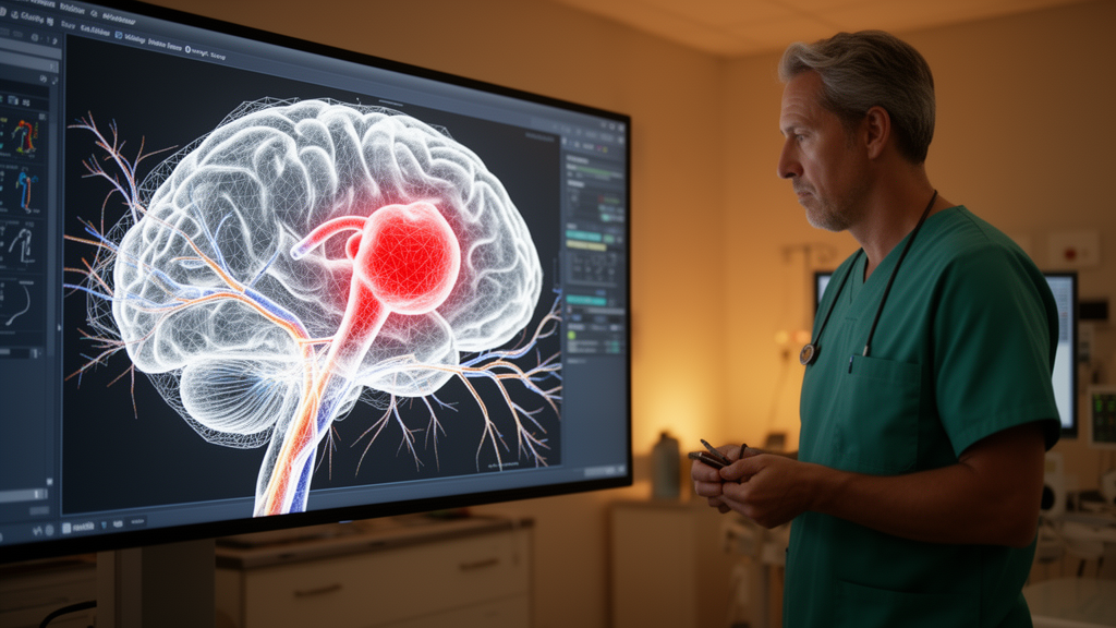
Deep learning tools for cerebrovascular care show real promise but also stubborn gaps, according to a systematic review published in 3D Printing in Medicine. Following PRISMA guidelines, researchers searched PubMed, IEEE Xplore, Scopus and Web of Science for peer-reviewed studies from 2015 to 2026 and screened 1,847 records, of which 127 met their inclusion criteria for deep learning applied to cerebrovascular image segmentation, multimodal fusion, hemodynamic prediction or 3D printing.
The review’s first key finding concerns segmentation. Three-dimensional U-Net architectures equipped with attention mechanisms achieved state-of-the-art performance for major cerebral vessels, with Dice coefficients ranging from 0.85 to 0.92. However, the authors report that performance degrades significantly for vessels smaller than 0.5 millimeters, a limitation that matters because fine vessels are often central to treatment decisions in intracranial aneurysm care.
On the imaging side, the review traces how multimodal fusion strategies have evolved from early and late fusion toward hybrid architectures incorporating transformers and cross-attention mechanisms. These newer designs improved fusion fidelity by 15 to 25 percent over unimodal approaches, suggesting that combining information from different imaging modalities through learned attention can outperform single-source pipelines.
Hemodynamic modeling is the third pillar. The review highlights reduced-order modeling that combines Proper Orthogonal Decomposition with LSTM networks, enabling real-time hemodynamic prediction while preserving more than 99 percent of flow energy with fewer than ten modes. This kind of compression could make patient-specific flow simulation fast enough for clinical workflows, though the review frames this as methodological potential rather than established practice.
Materials remain a weak point for 3D printing. Biomechanical characterization summarized in the review indicates that silicone elastomers more closely match native vascular tissue, with elastic moduli of 0.02 to 1.16 newtons per square millimeter, than current 3D-printed materials, which fall between 0.85 and 1.02 newtons per square millimeter. Notably, the authors state that both material classes fail to replicate the nonlinear behavior of real tissue, so printed models still fall short of true mechanical fidelity.
Explainable AI is increasingly used in this field, with methods such as Grad-CAM, SHAP and LIME appearing across the included studies. Yet the review finds these approaches lack standardized validation protocols and clinical expert involvement in their evaluation, leaving a gap between technical interpretability and clinically meaningful transparency.
The context for this work is significant: cerebrovascular diseases, particularly intracranial aneurysms, are described in the review as a leading cause of mortality and long-term disability worldwide. The authors argue that integrating deep learning with multimodal imaging, hemodynamic modeling and 3D printing offers transformative potential for personalized neurosurgical planning, but that the field has lacked a comprehensive synthesis of methodological approaches, validation frameworks and clinical translation pathways, which this review set out to provide.
The review’s conclusion is measured. Deep learning-based multimodal integration has demonstrated significant potential for advancing cerebrovascular surgical planning, but critical gaps remain in small vessel segmentation, biomechanical material fidelity, standardized validation frameworks and prospective clinical validation. The authors recommend that future research prioritize foundation model development, physics-informed neural networks, multi-material 3D printing with programmable properties, and human-centered explainability evaluation to enable clinical translation.
Because this is a systematic review of prior studies rather than a new clinical trial, its findings describe the state of the literature rather than direct patient outcomes. The quality assessment was performed using tailored instruments for each methodological domain, and the identified performance ranges reflect the included studies. For clinicians and researchers, the message is that the individual components of an intelligent cerebrovascular intervention pipeline are advancing rapidly, but connecting them into validated, clinically deployable systems remains the field’s central unfinished task.
Subject of Research: A systematic review of deep learning methods for cerebrovascular image processing, hemodynamic modeling and 3D printing in surgical planning
Article Title: Intelligent cerebrovascular intervention: a deep learning approach to multimodal image processing, hemodynamic modeling, and 3D printing
Article References: Nirala, S., Hammad, A., Khan, N. U., & Huang, C. (2026). Intelligent cerebrovascular intervention: a deep learning approach to multimodal image processing, hemodynamic modeling, and 3D printing. 3D Printing in Medicine. https://doi.org/10.1186/s41205-026-00351-9
Image Credits: AI Generated
DOI: 10.1186/s41205-026-00351-9
Keywords: deep learning, intracranial aneurysm, image segmentation, multimodal fusion, hemodynamic modeling, 3D printing, systematic review, U-Net, reduced-order modeling, explainable AI, neurosurgical planning, Intelligent
Cite Scienmag News
Ophelia Keating. (October 27, 2026). Deep learning maps a path toward smarter aneurysm surgery planning. Scienmag. https://scienmag.com/deep-learning-maps-a-path-toward-smarter-aneurysm-surgery-planning/
Ophelia Keating. "Deep learning maps a path toward smarter aneurysm surgery planning." Scienmag, 27 October 2026, https://scienmag.com/deep-learning-maps-a-path-toward-smarter-aneurysm-surgery-planning/. Accessed 27 October 2026.
Ophelia Keating. "Deep learning maps a path toward smarter aneurysm surgery planning." Scienmag. October 27, 2026. https://scienmag.com/deep-learning-maps-a-path-toward-smarter-aneurysm-surgery-planning/


