


People with type 2 diabetes can lose the ability to distinguish fine shades of gray before any diabetic retinopathy shows up in the eye, and a new study suggests that this early functional decline goes hand in hand with measurable thinning of a specific retinal layer. The research, published in Frontiers in Medicine, examined 90 Chinese adults aged 40 to 60 years and found that contrast sensitivity — the visual system’s ability to detect objects against their background — was already reduced in people with diabetes who had no clinically detectable retinopathy, and reduced further in those with mild nonproliferative diabetic retinopathy.
The study, a prospective cross-sectional investigation conducted at Lixiang Eye Hospital of Soochow University between January 2023 and December 2025, divided participants into three groups of 30 each: healthy controls, individuals with type 2 diabetes without diabetic retinopathy, and individuals with diabetes and mild nonproliferative diabetic retinopathy. All participants were recruited from the ophthalmology outpatient clinic, and the researchers excluded anyone with conditions that could compromise vision, including cataracts, glaucoma, diabetic macular edema, retinal detachment, or vitreous hemorrhage, as well as people with proliferative diabetic retinopathy or severe systemic diseases.
Contrast sensitivity was assessed with the CSV-1000E chart, which tests the ability to detect sine-wave gratings at four fixed spatial frequencies: 3, 6, 12, and 18 cycles per degree. Testing was performed monocularly at 2.5 meters under standardized photopic conditions, and the same trained examiner, blinded to participants’ diabetic status and clinical grouping, conducted all examinations. The researchers summarized overall contrast sensitivity using the area under the log contrast sensitivity function, known as AULCSF, and classified low-, middle-, and high-spatial-frequency sensitivity as corresponding to 3, 6–12, and 18 cycles per degree, respectively.
The results showed that AULCSF and middle-spatial-frequency contrast sensitivity differed significantly across the three groups, while low- and high-spatial-frequency sensitivity did not. Post-hoc analyses revealed that AULCSF was significantly lower in every pairwise comparison among the groups. Middle-spatial-frequency contrast sensitivity was significantly lower in both the no-retinopathy and nonproliferative retinopathy groups than in controls, although the difference between the two diabetes groups did not survive Bonferroni correction. Median AULCSF fell from 1.13 in controls to 1.05 in the no-retinopathy group and 0.91 in the nonproliferative retinopathy group.
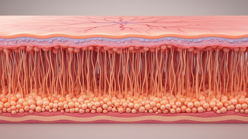
The structural counterpart to this functional decline emerged from optical coherence tomography imaging. Using a TowardPi swept-source OCT system with an axial resolution of 6 micrometers or less, the team measured photoreceptor layer thickness, defined as the vertical distance between the external limiting membrane and the inner boundary of the retinal pigment epithelium. This composite measurement includes the photoreceptor inner and outer segment regions but excludes the outer nuclear layer and the retinal pigment epithelium–Bruch’s membrane complex, so the researchers interpreted it as thinning of a photoreceptor-related compartment rather than direct evidence of photoreceptor cell loss.
Photoreceptor layer thickness showed a progressive reduction across diabetic stages: median values fell from 30.4 micrometers in controls to 27.4 micrometers in the no-retinopathy group and 25.4 micrometers in the nonproliferative retinopathy group, with Bonferroni-adjusted post-hoc tests confirming significant differences across all three pairwise comparisons. In contrast, ganglion cell–inner plexiform layer thickness was significantly lower only in the nonproliferative retinopathy group, dropping to 83.5 micrometers compared with roughly 87 micrometers in the other two groups. Total, inner, and outer retinal thickness showed no significant differences among the groups.
The vascular story was notably quieter. Foveal avascular zone area, vessel density, and amplitude-decorrelation Fourier-domain imaging values of the superficial and deep capillary plexuses showed no statistically significant differences among the three groups. The authors caution that the absence of significant differences does not exclude subtle or early microvascular alterations, particularly given the sample size and cross-sectional design, and they note that whether neuroretinal structural and functional changes precede microvascular changes requires investigation in longitudinal studies.
The central finding of the study was the link between structure and function. Photoreceptor layer thickness was positively correlated with middle-spatial-frequency contrast sensitivity across all participants (r = 0.385, P = 0.0024), an association that remained significant after Benjamini–Hochberg false discovery rate correction and in a Spearman sensitivity analysis. The association also persisted in multivariable regression among participants with diabetes after adjustment for age, sex, axial length, type 2 diabetes duration, and glycated hemoglobin, and it remained significant in an extended sensitivity model that additionally accounted for best-corrected visual acuity, hypertension, and hyperlipidemia. Exploratory subgroup analyses found the correlation was significant only within the nonproliferative retinopathy group (r = 0.572, P = 0.001), not among controls or the no-retinopathy group.
The researchers also explored whether photoreceptor layer thickness could identify people with abnormal contrast sensitivity among the 60 participants with diabetes. Using a control-derived threshold — the empirical fifth percentile of the control group’s middle-spatial-frequency distribution, corresponding to a cutoff of 0.8235 — 16 of the 60 participants with diabetes were classified as having abnormal contrast sensitivity. Photoreceptor layer thickness differed significantly between those with normal and abnormal sensitivity, and exploratory ROC analysis yielded an area under the curve of 0.920 (95% confidence interval: 0.852–0.989), with an optimal cutoff of 25.45 micrometers giving 93.8% sensitivity and 86.4% specificity. The authors stress that this threshold was internally derived from the same small, single-center cohort, that the estimated performance may be optimistic, and that the analysis reflects the ability of a structural parameter to distinguish a functional test-defined outcome rather than a capacity to diagnose diabetic retinopathy or predict future visual impairment.
Among systemic inflammatory markers, only the platelet-to-lymphocyte ratio differed significantly among the three groups, with lower values in the nonproliferative retinopathy group than in the other two. This ratio showed a weaker positive correlation with middle-spatial-frequency contrast sensitivity that did not survive multiple-testing correction, so the authors treat any link between systemic inflammation and retinal function as exploratory. The study has other limitations the authors acknowledge: the sample was small, all participants were Chinese, photoreceptor thickness was manually measured by a single examiner without formal repeatability assessment, the examiner’s masking status was not documented, and exploratory cell density and cell regularity indices could not be fully verified. Given the cross-sectional design, the findings should be read as associations rather than evidence of causality, and longitudinal studies are needed to determine whether these early structural and functional changes predict the later development and progression of diabetic retinopathy.
Subject of Research: Early retinal structural and contrast sensitivity changes in type 2 diabetes without clinically detectable diabetic retinopathy
Article Title: Reduced photoreceptor layer thickness in early type 2 diabetes mellitus is associated with decreased contrast sensitivity: a prospective cross-sectional study
Article References: Tu, Y., Zhu, X., Sun, Y., Sun, X., & Cao, G. (2026). Reduced photoreceptor layer thickness in early type 2 diabetes mellitus is associated with decreased contrast sensitivity: a prospective cross-sectional study. Frontiers in Medicine, 13, Article 1984894. https://doi.org/10.3389/fmed.2026.1984894
Image Credits: AI Generated
DOI: 10.3389/fmed.2026.1984894
Keywords: type 2 diabetes, diabetic retinopathy, contrast sensitivity, photoreceptor layer, optical coherence tomography, retina, OCT angiography, nonproliferative diabetic retinopathy, retinal thickness, visual function, cross-sectional study, ophthalmology
Cite Scienmag News
Ophelia Keating. (October 27, 2026). Early Diabetes Thins a Light-Sensing Retinal Layer and Dulls Contrast Vision. Scienmag. https://scienmag.com/early-diabetes-thins-a-light-sensing-retinal-layer-and-dulls-contrast-vision/
Ophelia Keating. "Early Diabetes Thins a Light-Sensing Retinal Layer and Dulls Contrast Vision." Scienmag, 27 October 2026, https://scienmag.com/early-diabetes-thins-a-light-sensing-retinal-layer-and-dulls-contrast-vision/. Accessed 27 October 2026.
Ophelia Keating. "Early Diabetes Thins a Light-Sensing Retinal Layer and Dulls Contrast Vision." Scienmag. October 27, 2026. https://scienmag.com/early-diabetes-thins-a-light-sensing-retinal-layer-and-dulls-contrast-vision/


