


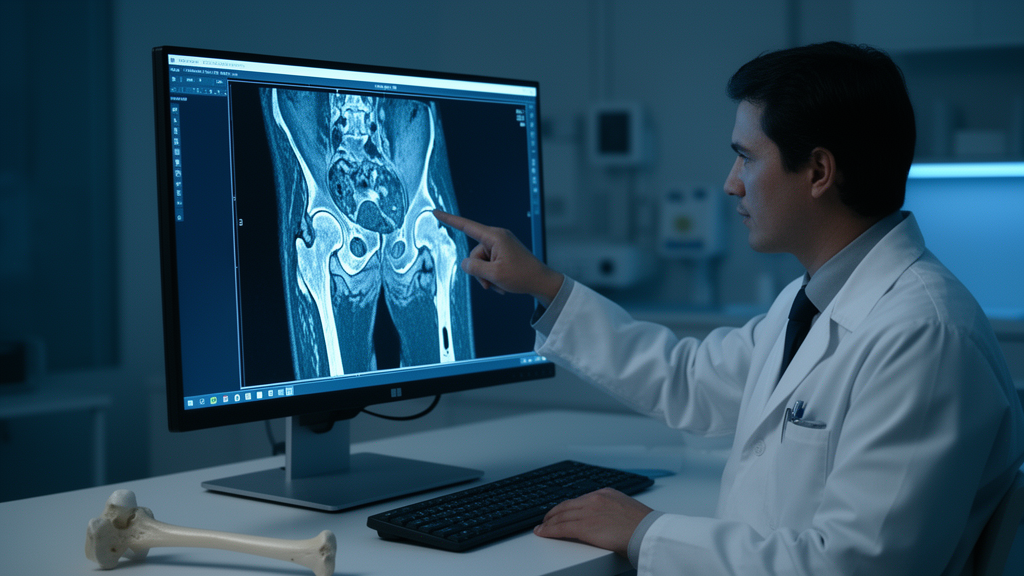
A single MRI feature visible just three months after surgery for a broken femoral neck may reveal, with striking consistency, which patients will go on to develop collapse of the femoral head, according to a retrospective cohort study published in Frontiers in Medicine. Researchers in China followed 148 patients with traumatic osteonecrosis of the femoral head (TONFH) and found that a repair band extending to the medial side of the femoral head, marked against the fovea of the ligamentum teres on coronal MRI, independently predicted collapse with an odds ratio of 13.350. The finding, if validated in larger multicenter cohorts, could give orthopedic surgeons an early imaging warning sign at a stage when intervention may still delay or even prevent the loss of hip joint function.
TONFH is one of the most feared complications of femoral neck fracture. The injury disrupts the blood vessels supplying the femoral head, and when that supply is inadequate, bone tissue dies. Once avascular necrosis progresses to collapse of the articular surface, patients typically lose hip function and often require total hip arthroplasty.
Identifying which fractures are destined to collapse before the collapse occurs has therefore been a persistent goal in orthopedic trauma research. Existing tools for assessing osteonecrosis arrive late in the disease course. The ARCO staging system grades osteonecrosis by the degree of femoral head collapse, and the JIC classification evaluates the size and location of the necrotic lesion, but both are applied only after osteonecrosis has already developed.
For patients with femoral neck fractures, clinicians want a way to predict TONFH before it happens, because that knowledge would change treatment decisions. MRI is recognized as the gold standard for diagnosing osteonecrosis, and prior work by Kawasaki and colleagues showed that early MRI after cannulated screw fixation can effectively indicate the presence of osteonecrosis. Advances in screw metal composition and metal artifact reduction sequences have also mitigated the image degradation that once limited MRI around implanted hardware.
The new study focuses on a specific MRI signature: the repair band. On T1-weighted images it appears as a low-signal band, and on T2-weighted images it produces the classic double-line sign. Pathologically, the band represents the transition zone between dead bone and newly formed bone, a sclerotic rim created as the body attempts to biologically seal the necrotic focus through local bone remodeling.
This structure is a double-edged sword. It can contribute to stress distribution across the femoral head, but it may also impede blood supply from reaching the surviving bone. When bone remodeling falls out of balance, micro-fractures accumulate and can extend to the subchondral bone plate, ultimately producing subchondral collapse under load.
What makes traumatic osteonecrosis distinct is its vascular origin. Non-traumatic ONFH is driven by systemic factors and produces necrotic lesions distributed across the medial, central, and lateral regions of the femoral head, so the location of the repair zone varies with the site of the lesion. In TONFH, by contrast, the injury damages the lateral epiphyseal arteries, and the resulting necrosis predominantly involves the lateral weight-bearing region, with isolated medial or central involvement being rare.
The distribution of repair zones therefore undergoes characteristic alterations in trauma patients, which is precisely why the research team hypothesized that the position of the repair band on MRI could serve as a risk marker for collapse specific to TONFH. The study was a single-center retrospective cohort conducted at Quanzhou Orthopedic-Traumatological Hospital, approved by the institutional review board, with all participants providing informed consent. The researchers screened 374 patients treated with cannulated screw fixation for femoral neck fracture between January 2016 and May 2023.
After excluding patients with incomplete data, ages outside 18 to 60 years, concomitant hip diseases such as rheumatoid or gouty arthritis, non-traumatic or pathological fractures, loss to follow-up, absence of TONFH at final follow-up, or follow-up shorter than two years, 148 patients with 148 affected hips remained. All surgeries were performed by the same team using closed reduction and three cannulated screws placed in an inverted triangular configuration, with weight-bearing initiated at three months and MRI performed uniformly at that same three-month postoperative mark. The cohort had a median age of 46.50 years, with 67 men and 81 women, and a median follow-up of 44.50 months.
Collapse, defined as a loss of femoral head height exceeding 2 millimeters on anteroposterior radiographs compared with the earliest post-reduction films, occurred in 63 hips, while 85 hips remained intact. The 2-millimeter threshold was chosen because previous studies have shown that collapse beyond this degree is significantly associated with worsening symptoms and the need for secondary surgery. Two senior surgeons independently assessed the imaging, and the interobserver intraclass correlation coefficient for identifying the medial repair band was 0.802, which the authors describe as excellent reliability.
Age, sex, affected side, achievement of anatomical reduction, and performance of emergency surgery showed no statistically significant differences between the collapse and non-collapse groups. The association between repair band position and outcome was stark. A medial repair band was present in 84.13 percent of the collapse group but only 27.06 percent of the non-collapse group, a difference with a P-value below 0.001.
In univariate logistic regression, complete fracture displacement, corresponding to Garden type IV, carried an odds ratio of 2.208 for collapse, while the medial repair band carried an odds ratio of 14.287. When both variables entered a multivariate model, the medial repair band remained an independent risk factor with an odds ratio of 13.350 and a 95 percent confidence interval of 5.767 to 30.907, whereas complete displacement lost its independent significance. Kaplan-Meier survival analysis confirmed that patients with a medial repair band had significantly lower femoral head survival than those with a lateral band, with median times to collapse of 27.00 months versus 29.50 months.
The study also probed why some repair bands extend medially. Using the fovea of the ligamentum teres as an anatomical landmark, the researchers linked band position to the vascular anatomy of the femoral head, particularly the perfusion territories of the superior retinacular artery and the inferior retinacular artery, whose anastomotic site lies adjacent to the fovea. In incompletely displaced fractures of Garden types I through III, the inferior retinacular artery often maintains compensatory capacity, limiting the necrotic area so that the repair band terminates lateral to the fovea.
In completely displaced Garden type IV fractures, both arterial systems are severely damaged, producing extensive necrosis and a repair band that extends medially. After adjusting for reduction quality and surgical timing, complete displacement increased the risk of medial repair band formation by approximately 2.58-fold, with an odds ratio of 2.581 and a confidence interval of 1.304 to 5.106. Supporting evidence from prior research reinforces this vascular interpretation.
Lee and colleagues followed 33 cases of focal traumatic osteonecrosis confined to the lateral area for six years and observed no collapse, consistent with the favorable outcomes seen in the lateral repair band group here. Boraiah and colleagues used gadolinium-enhanced MRI to quantify blood supply and found that the inferior retinacular artery can contribute up to 20 percent of total femoral head perfusion, while Zhao and colleagues demonstrated through micro-CT and digital subtraction angiography that the inferior retinacular artery system serves as an important structure preserving blood supply after femoral neck fracture. The authors also note that some scholars have suggested Garden type IV fractures may contain subtypes, since outcomes vary considerably within that group, and the medial-versus-lateral repair band distinction offers new imaging-based evidence for such subclassification.
The authors propose that assessing the extent of the repair band on coronal MRI, using the ligamentum teres fovea as a landmark, provides a simple and reproducible method for gauging collapse risk in TONFH, with relatively high accuracy. A medial band should be considered a key early warning sign of impending collapse, while a lateral-only band suggests preserved compensatory perfusion and robust reparative potential. The study carries important limitations, however.
Its retrospective design introduces selection bias and loss to follow-up, the single-center sample is limited, only measurable confounders could be adjusted for, axial and sagittal MRI sequences were not routinely included, and because MRI was performed at a single three-month time point, the dynamic evolution of the repair band could not be captured. The authors call for larger, multicenter studies with long-term follow-up to validate the findings before this imaging sign becomes a routine clinical decision tool.
Subject of Research: MRI medial repair band as a predictor of femoral head collapse in traumatic osteonecrosis after femoral neck fracture
Article Title: Medial repair band: independent risk factor for collapse in traumatic osteonecrosis of the femoral head
Article References: Wang, Y., Huang, C., Gong, Z., Li, Z., He, W., Wu, Z., & Zhuang, Z. (2026). Medial repair band: independent risk factor for collapse in traumatic osteonecrosis of the femoral head. Frontiers in Medicine, 13, Article 2001241. https://doi.org/10.3389/fmed.2026.2001241
Image Credits: AI Generated
DOI: 10.3389/fmed.2026.2001241
Keywords: traumatic osteonecrosis of the femoral head, femoral neck fracture, MRI repair band, femoral head collapse, Garden classification, inferior retinacular artery, cannulated screw fixation, ARCO staging, orthopedic trauma, retrospective cohort study, hip joint preservation, metal artifact reduction sequences
Cite Scienmag News
Ophelia Keating. (October 26, 2026). MRI Sign Spots Which Broken Hips Will Collapse After Femoral Neck Fractures. Scienmag. https://scienmag.com/mri-sign-spots-which-broken-hips-will-collapse-after-femoral-neck-fractures/
Ophelia Keating. "MRI Sign Spots Which Broken Hips Will Collapse After Femoral Neck Fractures." Scienmag, 26 October 2026, https://scienmag.com/mri-sign-spots-which-broken-hips-will-collapse-after-femoral-neck-fractures/. Accessed 26 October 2026.
Ophelia Keating. "MRI Sign Spots Which Broken Hips Will Collapse After Femoral Neck Fractures." Scienmag. October 26, 2026. https://scienmag.com/mri-sign-spots-which-broken-hips-will-collapse-after-femoral-neck-fractures/


