


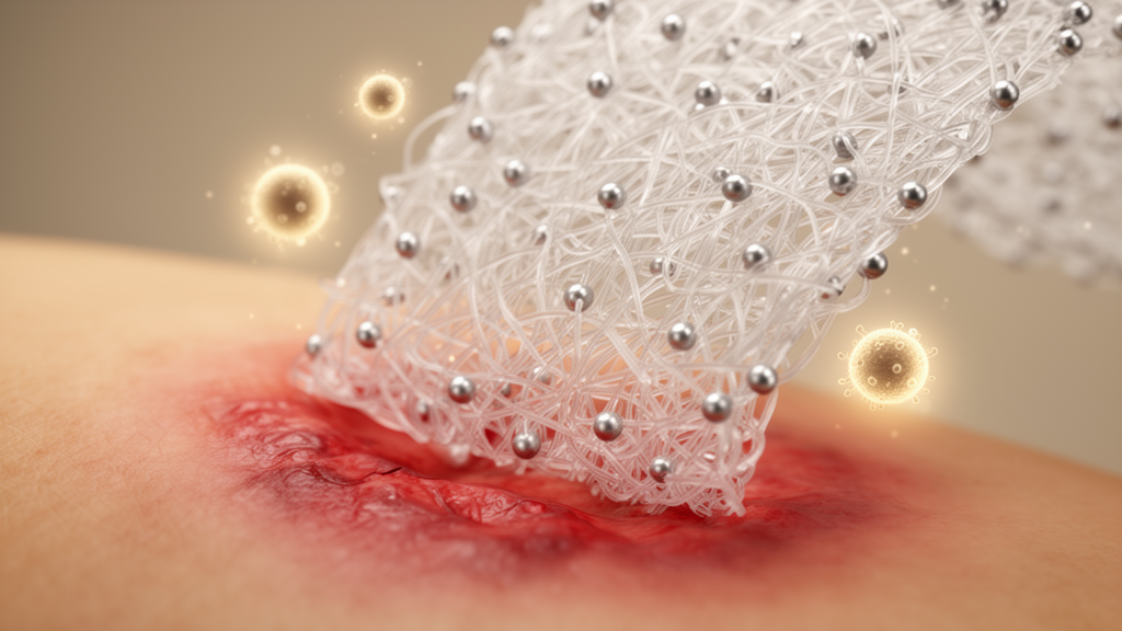
A new antibacterial wound dressing built from two industrially proven manufacturing techniques has shown potent activity against two of the most stubborn wound pathogens, methicillin-resistant Staphylococcus aureus (MRSA) and Pseudomonas aeruginosa, while releasing no detectable silver nanoparticles under the tested conditions. The dressing, described in a new materials study, combines flame spray pyrolysis with electrospinning to produce a nanofibrous membrane in which ultra-small silver nanoparticles are embedded inside fibers made of polyvinyl alcohol and chitosan. In laboratory tests, membranes containing the highest chitosan fractions reduced bacterial colony counts by 8 to 10 orders of magnitude, and the system achieved measurable antibacterial effects even when applied to infected samples of freshly excised human skin.
The motivation behind the work is the growing difficulty of treating wound infections with conventional antibiotics. Acute wound infections are particularly prevalent in burns, surgical sites, and traumatic wounds, and the primary pathogens are gram-positive MRSA and gram-negative P. aeruginosa. According to the study, approximately 70 percent of bacteria causing wound infections have become resistant to at least one antibiotic, contributing to more than 700,000 deaths annually. Existing alternatives include surgical debridement, which carries a significant risk of reinfection, and commercial antimicrobial dressings containing antiseptics such as iodine or chlorhexidine acetate, or ionic and nano-crystalline silver. Silver itself has long been valued because its multiple antibacterial mechanisms make the development of bacterial resistance less likely, but concerns about toxicity, inconsistent evidence of efficacy, and cost-effectiveness have limited its wider deployment.
The researchers addressed these limitations by choosing manufacturing routes that are already established at industrial scale. Flame spray pyrolysis produced multi-component Ag/SiO2 nanoparticles containing 50 percent silver by weight, in which amorphous silica inhibits the growth of silver during synthesis and prevents the particles from agglomerating in aqueous environments. Electron microscopy of the resulting particles showed a lognormal size distribution with a mean diameter of 4.7 nanometers, a sub-10-nm average size that the study identifies as critical for enabling high silver ion release. Selected-area electron diffraction and X-ray diffraction confirmed the crystallinity of the silver, with the average crystal size measured by Rietveld analysis coming out at approximately 12 nanometers, a volume-weighted figure that is disproportionately influenced by a few larger crystalline particles compared with the number-weighted distribution obtained from transmission electron microscopy.
These nanoparticles were then incorporated into a polymer solution and electrospun into nanofibers. The polymers polyvinyl alcohol and chitosan were selected for their biocompatibility and suitability for electrospinning, and both dissolve in aqueous solvents, which prevented the precipitation of silver nanoparticles and created an environment compatible with future incorporation of biomolecules. The solvent was 2 percent acetic acid, needed for chitosan dissolution, with Tween 20 acting as a surfactant and glycerol as a plasticizer. Membranes were fabricated with chitosan contents ranging from 0 to 20 percent of the total polymer weight while keeping the nanoparticle amount constant, allowing a systematic evaluation of how composition affects antibacterial activity. As chitosan content increased, the viscosity of the polymer solution rose, requiring higher voltage for electrospinning and yielding thinner fibers, but all membranes retained fiber diameters within 150 to 300 nanometers and a porosity of around 40 percent. Electron tomography and elemental mapping confirmed a homogeneous distribution of the Ag/SiO2 nanoparticles across the fibers at all chitosan concentrations.
The behavior of the membranes in water revealed the central design principle of the system. Fibers made entirely of polyvinyl alcohol dissolved rapidly on contact with water, fully releasing the incorporated Ag/SiO2 nanoparticles, which ultraviolet-visible spectroscopy confirmed through a surface plasmon resonance peak at 420 nanometers. In contrast, no nanoparticle release was detected by ultraviolet-visible spectroscopy from chitosan-containing fibers over the tested 24-hour period, indicating that chitosan prevents polyvinyl alcohol dissolution and traps the nanoparticles inside the polymer matrix. Despite this retention, the membranes with 10 and 20 percent chitosan showed the highest sustained release of silver ions, enabled by the ultra-small size of the embedded silver particles. Inductively coupled plasma measurements showed that the silver content is remarkably low: 1 milligram of membrane contains just 20 micrograms of silver, meaning a 1-square-centimeter membrane carries approximately 20 micrograms. The high ion release from this minimal loading is attributed to the dissolution of the intrinsic silver oxide layer on the nanoparticle surface, an effect pronounced in the higher-chitosan membranes.
Detailed physicochemical characterization explained why chitosan plays this dual role. Both polymers are hydrophilic and absorb water, and the resulting swelling creates more space for silver ions to diffuse out of the Ag/SiO2 nanoparticles. Chitosan is derived from the deacetylation of naturally occurring chitin, with a degree of deacetylation of 75 to 85 percent, leaving residual acetyl groups that undergo hydrolysis in aqueous environments to form acetic acid and lower the local pH. Silica enhances this hydrolysis, further reducing the pH in membranes containing the nanoparticles. Increased acidity promotes silver dissolution through a redox reaction that releases silver ions, and as the pH decreases, the amine groups of chitosan become protonated, causing electrostatic repulsion between polymer chains and additional swelling. This accounts for the higher swelling ratio of 200 percent observed in membranes with 20 percent chitosan compared with 100 percent in those with 10 percent. The study also noted that the high voltages of 10 to 18 kilovolts used in electrospinning did not alter the oxide-to-metallic silver ratio in the membranes, as measured by X-ray photoelectron spectroscopy, leaving the ion release profiles unaffected.
Antibacterial testing against MRSA and P. aeruginosa produced striking results in vitro. After 18 hours of incubation, membranes containing 10 and 20 percent chitosan achieved 8-log and 10-log reductions in bacterial colony-forming units, respectively, exceeding 99.9999 percent. The effect was notably stronger against P. aeruginosa than MRSA, a difference the study attributes to bacterial cell structure. Gram-positive MRSA has a thick peptidoglycan layer of approximately 30 nanometers that renders it less vulnerable to penetration by silver ions, whereas gram-negative P. aeruginosa has a much thinner peptidoglycan layer of 3 to 4 nanometers and possesses negatively charged lipopolysaccharides in the cell membrane that promote the adhesion of silver ions. Microscopy of MRSA cells after incubation showed morphology characteristic of damaged cells. A disk diffusion assay demonstrated a clear inhibition zone around the membranes, indicating that the antibacterial effect comes from the high diffusivity of released silver ions rather than direct contact with the membranes or diffused nanoparticles. When the researchers substituted silver nitrate for the Ag/SiO2 nanoparticles at matched silver content, the resulting membranes showed slightly lower ion release and comparable antibacterial activity, but chitosan in the polymer matrix reduced the silver nitrate to silver nanoparticles upon contact with water, raising toxicity concerns and favoring the original nanoparticle design.
The system was then evaluated in more demanding ex vivo models. Wounds created on thawed porcine skin were infected with 105 colony-forming units per wound, allowed to colonize for 2 hours, and treated for 4 hours; the membrane with 20 percent chitosan achieved a 3-log reduction, or 99.9 percent, in MRSA colony counts even in this growth-conducive environment. On freshly excised human skin from plastic surgery donors, the same membrane achieved a 1-log reduction, or 90 percent, against MRSA and a 2-log reduction, or 99 percent, against P. aeruginosa. The study notes that human skin varies considerably in age, gender, anatomical location, and biological factors, which accounts for the inter-donor and intra-donor variability observed. Testing on ex vivo human skin is considered more physiologically relevant than porcine or rodent models, and the authors describe these experiments as initial evidence of antibacterial activity in human skin tissue supporting further preclinical evaluation.
Biocompatibility testing addressed the toxicity concerns that have hindered many silver-based dressings. Histological analysis of human skin biopsies treated for 18 hours revealed no pathological changes, with tissue showing normal architecture including a well-differentiated epidermis and dense dermal connective tissue. For the chitosan-free membrane, released nanoparticles were visible on the wound surface, but no nanoparticle release was detected from the 20 percent chitosan membrane under the tested conditions, which the study suggests may reduce nanoparticle accumulation and mitigate associated long-term toxicity concerns. Immunohistochemistry showed low numbers of cleaved caspase-3 positive cells, an apoptosis marker, with proliferating cell nuclear antigen concentrated in the epithelium, both characteristic of healthy tissue. Membrane extracts prepared according to the ISO 10993-12 protocol were tested against primary human adult epidermal keratinocytes and dermal fibroblasts using a lactate dehydrogenase assay, and all experimental groups exhibited approximately 90 percent cell viability after 72 hours, indicating no substantial cytotoxicity under the tested conditions.
In wound-healing experiments on fresh human skin biopsies, the electrospun membranes performed comparably to untreated skin in assessed parameters of tissue contraction, re-epithelialization, and wound closure, with histology showing proliferating keratinocytes and activated fibroblasts characteristic of healing tissue. Under the tested ex vivo conditions, a commercially available silver-based dressing was associated with reduced wound-healing responses, including a lack of tissue contraction and re-epithelialization, and exhibited cytotoxicity toward fibroblasts despite having antibacterial activity only slightly higher than the electrospun membrane. The authors emphasize that the study was designed as a focused evaluation of antibacterial performance, nanoparticle retention, ion release behavior, cytocompatibility, and short-term wound-healing compatibility rather than a complete preclinical assessment. Long-term toxicity, inflammatory profiling, collagen deposition, angiogenesis, extended ion release, and comprehensive mechanical characterization remain subjects for future in vivo studies. The novelty of the work, the researchers argue, lies not in electrospinning or silver dressings themselves but in integrating flame-made, silica-stabilized sub-10-nm nanoparticles into a polyvinyl alcohol-chitosan matrix that enables sustained silver ion release at very low silver loading without detectable nanoparticle release, providing a basis for subsequent in vivo and preclinical evaluation.
Subject of Research: Antibacterial electrospun wound dressings with embedded flame-made silver nanoparticles for resistant wound infections
Article Title: Chitosan-stabilized electrospun nanofiber dressings with embedded flame-made nanosilver for the treatment of resistant infections
Article References: Ramachandran, R. V., Geara Semaan, J., Samara, M., Thersleff, T., Fielden, M., Sommar, P., Xu Landén, N., & Sotiriou, G. A. (2026). Chitosan-stabilized electrospun nanofiber dressings with embedded flame-made nanosilver for the treatment of resistant infections. iScience, 29(11), Article 117991. https://doi.org/10.1016/j.isci.2026.117991
Image Credits: AI Generated
DOI: 10.1016/j.isci.2026.117991
Keywords: wound dressings, silver nanoparticles, chitosan, electrospinning, flame spray pyrolysis, MRSA, Pseudomonas aeruginosa, antibiotic resistance, silver ion release, biocompatibility, ex vivo human skin, nanofibers
Cite Scienmag News
Denise Maddox. (October 24, 2026). Flame-made nanosilver woven into chitosan fibers fights resistant wound bacteria. Scienmag. https://scienmag.com/flame-made-nanosilver-woven-into-chitosan-fibers-fights-resistant-wound-bacteria/
Denise Maddox. "Flame-made nanosilver woven into chitosan fibers fights resistant wound bacteria." Scienmag, 24 October 2026, https://scienmag.com/flame-made-nanosilver-woven-into-chitosan-fibers-fights-resistant-wound-bacteria/. Accessed 24 October 2026.
Denise Maddox. "Flame-made nanosilver woven into chitosan fibers fights resistant wound bacteria." Scienmag. October 24, 2026. https://scienmag.com/flame-made-nanosilver-woven-into-chitosan-fibers-fights-resistant-wound-bacteria/


