


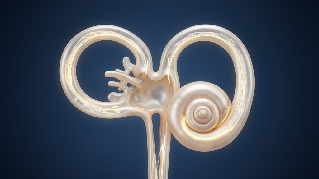
When a 55-year-old soldier woke up and sat upright in his bed one morning, the world began to spin so violently that he was nauseated and vomited. Lying flat made the dizziness recede; standing up brought it roaring back. Within hours he arrived at a neurological emergency department in Ljubljana, Slovenia, where clinicians observed something extraordinary: a relentless, direction-changing nystagmus, an involuntary jerking of the eyes, that flipped its horizontal direction depending on which way his head was turned while he lay on his back, and a persistent downward-beating component the moment he sat upright. The case, now published as a case report in Frontiers in Audiology and Otology by neurologists Lara Straus and Igor Rigler of the University Medical Center Ljubljana, may represent the first documented instance of an uncommon vestibular disorder, the light cupula phenomenon, affecting all three semicircular canals of a single inner ear simultaneously.
The light cupula phenomenon is a rare peripheral vestibular condition defined by a persistent, direction-changing positional nystagmus that appears rapidly, does not fatigue, and lacks the characteristic delay of benign paroxysmal positional vertigo, the far more common cause of positional dizziness. The prevailing hypothesis holds that the disorder arises when the density of the cupula, the gelatinous flap that seals each semicircular canal and senses fluid motion, falls below that of the surrounding endolymph, the fluid filling the canal. In that situation, gravity no longer leaves the cupula neutrally buoyant. Instead, it continuously deflects the sensory flap in whatever direction the head is oriented, producing a sustained nystagmus that changes direction as the head changes position, and vanishes only at a specific head angle known as the null plane.
Most reported cases of the light cupula phenomenon involve the horizontal semicircular canal, which lies roughly in the plane of the head when a patient is supine and is therefore the easiest canal to interrogate with positional testing. Involvement of the vertical canals, the anterior and posterior canals stacked in perpendicular planes above the ear, has been described only sparingly. Simultaneous involvement of all three canals within one vestibular apparatus has, according to the authors’ knowledge of the literature, not previously been documented. That is what makes the Slovenian soldier’s presentation so noteworthy: his eye movements suggested that the cupulae of his right horizontal, anterior, and posterior canals were all behaving as if they had become lighter than their surrounding endolymph at the same time.
The diagnostic reasoning rests on Ewald’s laws, nineteenth-century principles of vestibular physiology that remain central to interpreting nystagmus today. Ewald’s first law states that eye movements generated by a semicircular canal occur in that canal’s anatomical plane. Ewald’s second law holds that in the horizontal canal, deflection of the cupula toward the utricular cavity, the ampullopetal direction, produces a stronger excitatory response than deflection away from it. When the patient lay supine and his head was rotated 90 degrees to the right, the geotropic nystagmus, with its fast phase beating toward the ground, was markedly stronger than when the head was rotated to the left. That asymmetry localized the pathology to the right vestibular apparatus: turning the head to the left deflected the right horizontal cupula in the inhibitory, utriculofugal direction, yielding a weaker response.
Equally telling was the null plane. As the examiners rotated the patient’s head while he lay on his back, the nystagmus disappeared entirely when the head was turned approximately 15 degrees to the right of the vertical axis, then reversed direction when rotation continued further. The null plane is a critical sign in any patient with persistent direction-changing positional nystagmus, because it is typically present only in cupulopathies, disorders in which the cupula itself is abnormally deflected by gravity, and is generally absent in canalolithiasis, where freely floating otoconial debris triggers only brief, delayed, fatigable bursts of nystagmus. The authors attribute the slight rightward offset of the null plane in their patient to a small anatomical deviation of the horizontal canal cupula from the true vertical plane in the supine position.
The vertical component of the nystagmus demanded a more elaborate explanation. When seated, the patient immediately displayed a prominent downbeat nystagmus, with a large amplitude that did not diminish even after several minutes upright. According to Ewald’s third law, utriculofugal cupular deflection activates the sensory epithelium of both vertical canals. Because of the anatomical positioning of the ampullae in the vertical canals, a light cupula affecting both canals at once would deflect the anterior canal’s cupula in the excitatory utriculofugal direction while deflecting the posterior canal’s cupula in the inhibitory utriculopetal direction. The resulting pattern, anterior excitation paired with posterior inhibition, mimics the vestibulo-ocular reflex generated during cervical spine flexion, which drives a compensatory upward eye drift. The slow upward drift is then corrected by a downward saccade, producing the geotropic downbeating nystagmus the clinicians observed.
Strikingly, the nystagmus carried no torsional component, the rotational eye movement that vertical canal stimulation would ordinarily be expected to produce. The authors offer two hypotheses. Tilting the head to the right simultaneously activates both the right anterior and right posterior canals, which generate opposing torsional eye movements; under the light cupula alignment, anterior canal excitation and posterior canal inhibition may simply cancel each other’s torsional effects, leaving only the vertical component. Alternatively, observations from anterior canal benign paroxysmal positional vertigo suggest that the torsional component of anterior canal stimulation is not always as prominent as the vertical one, a pattern frequently described clinically. Either way, the coexistence of positional horizontal and vertical nystagmus without torsion fit the proposed simultaneous light cupula involvement of all three canals in the same ear.
Before settling on a peripheral explanation, the team worked hard to exclude central causes, which can also produce persistent direction-changing positional nystagmus. Brain computed tomography, computed tomography perfusion, computed tomography angiography, and later magnetic resonance imaging were all normal, as were blood tests. Neurological examination of the cranial nerves revealed no other abnormalities: the patient had no paresis, no ataxia, no hearing loss, no headache, and no sensory deficits. He could not walk unaided because of imbalance, but his gait showed no ataxia. Central disorders known to cause persistent geotropic positional nystagmus include cerebellar glioma, lateral medullary infarction, multiple sclerosis, and, more commonly, vestibular migraine, in which the nystagmus may persist for days; however, such central nystagmus is generally slow and lacks the null plane. With the null plane identified and no other neurological findings, the clinicians concluded the nystagmus most likely reflected a cupulopathy. The patient was referred to otorhinolaryngology, prescribed betahistine, and given balance exercises.
The clinical course was informative in its own right. The vertigo persisted for 14 days and ceased spontaneously, with gradual improvement: for the first 5 days the patient could not walk, and thereafter ambulation was possible only with support. Four months later a similar episode struck, with less intense vertigo but more pronounced nausea, lasting a week; an otorhinolaryngologist who examined him afterward found nothing remarkable. Over the following two and a half years he experienced only occasional brief episodes of vertigo lasting a few seconds, and at follow-up no neurological deficits were observed. This spontaneous resolution within roughly two weeks matches the natural history reported for most patients diagnosed with light cupula syndrome, and it also highlights a therapeutic dilemma: because the underlying etiology remains unclear, canalith repositioning maneuvers, the mainstay of treatment for canalolithiasis and cupulolithiasis, have limited effectiveness in the light cupula phenomenon.
Which mechanism rendered the cupulae light? The authors weigh three hypotheses. The lighter cupula theory proposes a decrease in cupular density, analogous to alcohol positional nystagmus, in which alcohol diffuses more rapidly into the cupula than into the endolymph because of the cupula’s proximity to capillaries; but that mechanism struggles to explain the syndrome’s acute onset. The heavier endolymph theory proposes an increase in endolymphatic density, potentially from acute inner ear injuries such as hypoperfusion, inflammation, or exudation; yet density changes in endolymph would be expected to affect all canals, which sits awkwardly with the predominantly horizontal canal involvement seen in clinical practice. The light debris theory presumes particles lighter than endolymph form and adhere to the cupula, possibly degenerated inflammatory cells or chemically altered otolith debris, which would fit the sudden onset, unilateral involvement, and gradual improvement, though the posterior canal, whose ampulla sits at the lowest point, should be less susceptible to debris with an upward settling tendency. Given the sudden onset and the joint disturbance of all three canals in their patient, the authors judge a disturbance of endolymph density due to hemorrhage or exudation affecting all three canals simultaneously to be the most plausible explanation.
The report carries important limitations, which the authors acknowledge candidly. Vertical nystagmus was observed only in an approximately upright head position; the head-bow and head-lean tests, which might have unmasked a torsional component or altered the vertical pattern, were not performed. No video-oculography or Frenzel glasses were used, so nystagmus was assessed solely by neurological examination, and no specialist assessment took place during the recurrent episode or in the long interval before follow-up. The precise anatomical positioning of the ampullae in the human vertical canals also remains poorly defined, and interindividual differences in their positions are far greater than in the horizontal canals, which could explain why patients with similar pathologies show different clinical findings. At this stage, the light cupula phenomenon in the vertical canals remains a plausible theoretical hypothesis rather than a settled diagnosis. The authors call for research with larger patient samples, systematic observation of nystagmus across different gravitational planes, and detailed study of vertical ampullary anatomy. For clinicians, the message is sharper: when persistent, direction-changing positional nystagmus presents with a null plane and an otherwise clean neurological examination, a cupulopathy deserves serious consideration, even when the pattern defies the textbook.
Subject of Research: A case report describing the light cupula phenomenon affecting all three semicircular canals of one vestibular apparatus
Article Title: Case Report: A light cupula phenomenon involving all three semicircular canals?
Article References: Straus, L., & Rigler, I. (2026). Case Report: A light cupula phenomenon involving all three semicircular canals?. Frontiers in Audiology and Otology, 4, Article 1988052. https://doi.org/10.3389/fauot.2026.1988052
Image Credits: AI Generated
DOI: 10.3389/fauot.2026.1988052
Keywords: light cupula, nystagmus, vertigo, vestibular, case report, semicircular canals, BPPV, neurology, Case, Report, light, cupula
Cite Scienmag News
Ophelia Keating. (October 23, 2026). Rare Inner Ear Disorder May Have Struck All Three Balance Canals at Once. Scienmag. https://scienmag.com/rare-inner-ear-disorder-may-have-struck-all-three-balance-canals-at-once/
Ophelia Keating. "Rare Inner Ear Disorder May Have Struck All Three Balance Canals at Once." Scienmag, 23 October 2026, https://scienmag.com/rare-inner-ear-disorder-may-have-struck-all-three-balance-canals-at-once/. Accessed 23 October 2026.
Ophelia Keating. "Rare Inner Ear Disorder May Have Struck All Three Balance Canals at Once." Scienmag. October 23, 2026. https://scienmag.com/rare-inner-ear-disorder-may-have-struck-all-three-balance-canals-at-once/


