


A combined model using high-frequency ultrasound (HFUS) and shear-wave elastography (SWE) identified children with Duchenne muscular dystrophy (DMD) more accurately than either imaging method alone in a retrospective study of 53 pediatric patients and 61 healthy controls. The combined approach achieved an area under the receiver operating characteristic curve (AUC) of 0.947, compared with 0.875 for HFUS and 0.846 for SWE. The study, published in Frontiers in Genetics, evaluated the methods as potential non-invasive adjuncts for assessing muscular involvement in DMD, rather than as replacements for genetic testing or biopsy.
Duchenne muscular dystrophy is an X-linked recessive disorder caused by deletions, duplications or point mutations in the dystrophin gene. Dystrophin is a core component of the muscle-fiber cytoskeleton, and its functional absence leads to repeated cycles of muscle-fiber necrosis and regeneration. Eventually, failed regeneration is accompanied by replacement with fibrotic tissue and fat infiltration, as well as involvement of the heart and respiratory muscles.
The study describes a global incidence of approximately one in 3,500 to 5,000 male live births and a prolonged disease course associated with severe functional decline, contractures and scoliosis in advanced stages. Diagnosis currently relies on clinical symptom assessment, serum muscle-enzyme assays, muscle biopsy and genetic testing. Genetic testing and muscle biopsy are regarded in the source article as diagnostic gold standards.
Genetic testing is particularly important because it can guide targeted therapies, but the investigators note that it is resource-intensive and may not be readily accessible as a first-line screening tool in every clinical setting. Muscle biopsy is technically challenging in children and, according to the study background, has low parental acceptance. The investigators reasoned that HFUS and SWE could provide complementary information.
HFUS was used to quantify muscle morphology, including thickness and cross-sectional area, while SWE measured tissue stiffness through shear-wave velocity. The study focused on the biceps brachii, a superficial upper-limb muscle, and the quadriceps femoris, a lower-limb group known from MRI and histological studies to undergo earlier and more severe fibrofatty change. Their superficial location also permits reproducible ultrasound imaging.
The researchers included 1,140 muscle sites and analyzed 3,420 parameters across 53 children with genetically or pathologically confirmed DMD and 61 age- and sex-matched healthy controls. Participants were recruited at West China Second University Hospital in China between January 2019 and September 2024. The control boys had normal physical examinations and no neuromuscular or other diseases.
Exclusion criteria included intellectual disability that prevented cooperation, current hormonal treatment or use within the previous six months, physical training for at least one month during the preceding three months, orthopedic surgery within the previous six months, and other conditions that clearly affected physical fitness. These restrictions reduced some sources of variation in muscle properties but also narrowed the population represented by the results. The groups did not differ significantly in age, weight or body-mass index, although children with DMD were shorter on average.
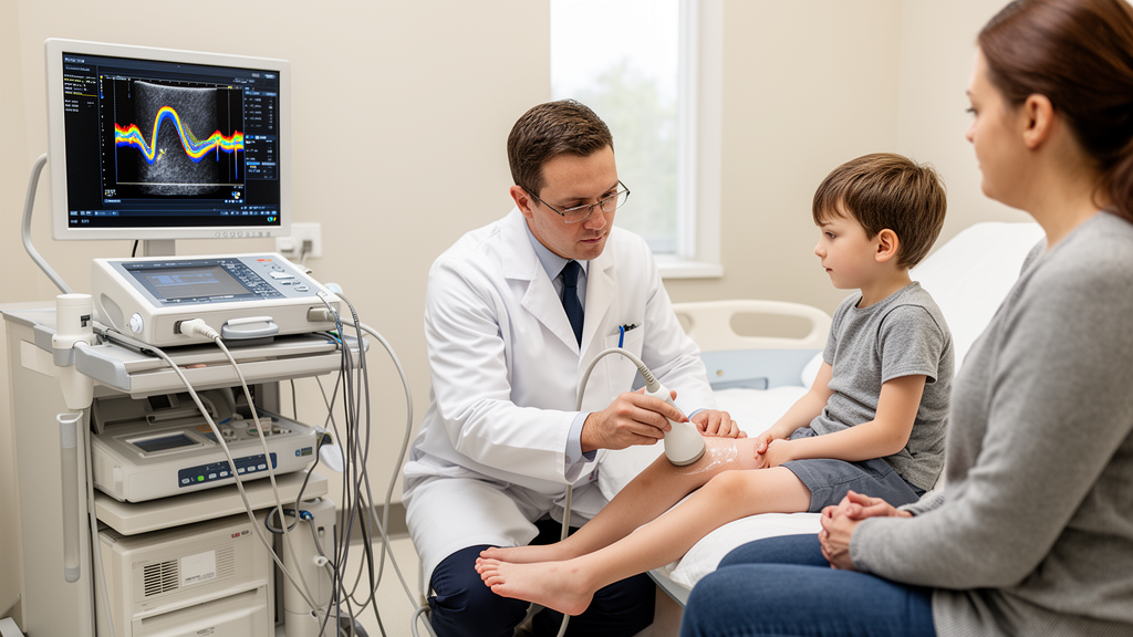
All examinations were performed with a Mindray Resona 7 system and a 5–14 MHz high-frequency linear-array probe. A certified sonographer with more than five years of experience in pediatric neuromuscular imaging examined the bilateral biceps brachii and quadriceps femoris while the children lay supine, relaxed, with their arms neutral and legs extended. HFUS was performed first, followed by SWE.
The sonographer assessed internal echogenicity, texture, blood-flow signals and space-occupying lesions, and measured muscle thickness and cross-sectional area. Mean shear-wave velocity, expressed in meters per second, was used to characterize stiffness. Standardized anatomical landmarks defined the measurement sites, and the entire muscle belly was included in cross-sectional-area measurements while surrounding fascia was excluded.
Two pediatric radiologists, each with more than 10 years of experience, independently analyzed the images without knowing participants’ clinical status or group allocation. Their measurements were averaged. Inter-observer reliability was generally high: intraclass correlation coefficients for muscle thickness, cross-sectional area and shear-wave velocity commonly ranged from 0.92 to 0.98.
The authors described these findings as evidence that the measurements were reproducible. For the HFUS model, principal component analysis reduced the number of morphological variables. The first four components had eigenvalues above 1, and the first two entered a multivariable logistic regression after univariate testing.
This model produced an AUC of 0.875, with a 95% confidence interval of 0.807 to 0.943, 88.9% sensitivity and 81.9% specificity. A Hosmer–Lemeshow test indicated good calibration. The separate SWE model was constructed through univariate and multivariable logistic regression.
Independent predictors included mean shear-wave velocity in the right biceps brachii, right vastus lateralis and left vastus intermedius. Their odds ratios were 0.170, 3.836 and 2.844, with 95% confidence intervals of 0.063 to 0.460, 1.623 to 9.070 and 1.002 to 8.075, respectively. The full SWE model achieved an AUC of 0.846, with a 95% confidence interval of 0.763 to 0.909.
The abstract reports 69.8% sensitivity and 75.4% specificity. A direct comparison found no statistically significant difference between the standalone HFUS and SWE models, with P = 0.494. Several individual measurements differed between the DMD and control groups.
Significant HFUS differences included selected thickness or cross-sectional-area measurements in the right biceps brachii and quadriceps muscles. Significant SWE differences occurred in the right biceps brachii, right vastus lateralis, left vastus medialis and left vastus intermedius. In every muscle with a significant SWE difference, shear-wave velocity was lower in the DMD group, indicating lower measured stiffness.
The authors interpret this pattern as consistent with the replacement of contractile muscle by softer adipose and connective tissue. They also note that the reduction differs from the increased stiffness reported in spastic conditions such as cerebral palsy. Left-right differences among homologous muscles were interpreted as compatible with an asymmetric and patchy disease process.
Combining the two models produced the study’s largest improvement in discrimination. The HFUS-SWE model reached an AUC of 0.947, with a 95% confidence interval of 0.885 to 0.981, and significantly outperformed both standalone models, with P < 0.001 for each comparison. Its sensitivity was 82.2%, while specificity reached 95.1%.
At the prevalence observed in this cohort, the combined model had a positive predictive value of 94.3% and a negative predictive value of 84.8%. Decision-curve analysis indicated a greater net benefit from the combined model across all tested threshold probabilities. The authors attribute its performance to complementary information: HFUS captures macroscopic morphology, including reduced muscle dimensions and architectural disruption, whereas SWE samples biomechanical properties such as tissue stiffness.
The findings remain preliminary because this was a small, single-center retrospective study, and the authors explicitly position the imaging approach as adjunctive. The cohort’s DMD prevalence was 46.5%, far above the global incidence of roughly one in 3,500 to 5,000 male births, so the reported predictive values cannot be assumed to apply to general screening populations. In a much lower-prevalence setting, the positive predictive value would be expected to be substantially lower and the negative predictive value higher.
Differences in height between the groups may also have influenced muscle biomechanics, although the authors argue that standardized measurement sites and relative ratios reduced this concern. Subcutaneous fat was considered as a possible confounder but could not be quantified because of its compressibility. Larger prospective, multicenter studies are needed to validate and refine the models, assess their performance in lower-prevalence populations and determine whether the approach can reliably support clinical triage or longitudinal monitoring.
Subject of Research: Evaluation of combined high-frequency ultrasound and shear-wave elastography models for adjunctive diagnosis of pediatric Duchenne muscular dystrophy.
Article Title: Non-invasive adjunctive diagnostic models incorporating high-frequency ultrasound and shear wave elastography for pediatric Duchenne muscular dystrophy
Article References: Qiang, K.-K., Zhang, S.-R., Luo, H., & Song, Q.-Y. (2026). Non-invasive adjunctive diagnostic models incorporating high-frequency ultrasound and shear wave elastography for pediatric Duchenne muscular dystrophy. Frontiers in Genetics, 17, Article 1912996. https://doi.org/10.3389/fgene.2026.1912996
Image Credits: AI Generated
DOI: 10.3389/fgene.2026.1912996
Keywords: Duchenne muscular dystrophy, Pediatric neuromuscular disease, High-frequency ultrasound, Shear-wave elastography, Diagnostic modeling, Muscle stiffness, Muscle morphology, Medical imaging, Non-invasive diagnosis, Non-invasive, adjunctive, diagnostic
Cite Scienmag News
Ophelia Keating. (October 21, 2026). Non-invasive adjunctive diagnostic models incorporating high-frequency ultrasound and shear wave elastography for pediatric Duchenne muscular dystrophy. Scienmag. https://scienmag.com/non-invasive-adjunctive-diagnostic-models-incorporating-high-frequency-ultrasound-and-shear-wave-elastography-for-pediatric-duchenne-muscular-dystrophy/
Ophelia Keating. "Non-invasive adjunctive diagnostic models incorporating high-frequency ultrasound and shear wave elastography for pediatric Duchenne muscular dystrophy." Scienmag, 21 October 2026, https://scienmag.com/non-invasive-adjunctive-diagnostic-models-incorporating-high-frequency-ultrasound-and-shear-wave-elastography-for-pediatric-duchenne-muscular-dystrophy/. Accessed 21 October 2026.
Ophelia Keating. "Non-invasive adjunctive diagnostic models incorporating high-frequency ultrasound and shear wave elastography for pediatric Duchenne muscular dystrophy." Scienmag. October 21, 2026. https://scienmag.com/non-invasive-adjunctive-diagnostic-models-incorporating-high-frequency-ultrasound-and-shear-wave-elastography-for-pediatric-duchenne-muscular-dystrophy/


