


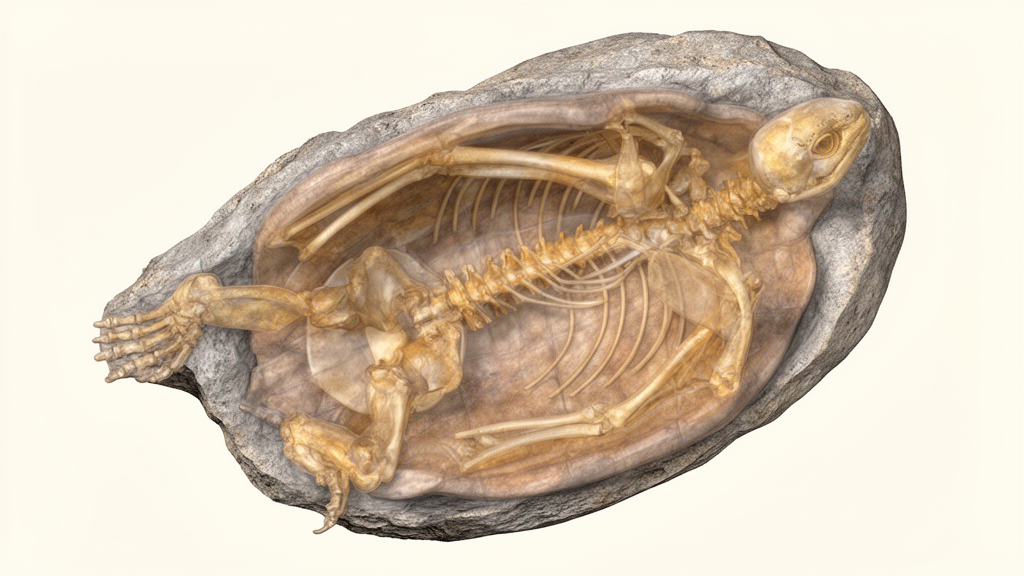
High-resolution X-ray micro-computed tomography and three-dimensional surface scanning have provided a detailed account of the skull and shell of a specimen of the Late Cretaceous turtle Denazinemys nodosa from southern Utah. The fossil, catalogued as DMNH EPV.64550, was recovered from the middle unit of the Kaiparowits Formation on the Kaiparowits Plateau in Grand Staircase-Escalante National Monument. It combines a largely complete skull with an associated, though disarticulated and partly incomplete, shell. The study used the scans to examine cranial sutures and internal canals, reconstruct the shell in three dimensions, revise the species diagnosis and investigate its relationships with other baenid turtles.
The locality is bracketed by dated volcanic ash beds and is approximately 76 million years old, placing the specimen in the late Campanian. The skull measures about 65 millimetres from the anterior tip of the nasals to the posterior end of the supraoccipital crest and 48 millimetres across the outer edges of the mandibular condyles. In dorsal view, it is wedge-shaped with a pinched snout, and the less-deformed side indicates dorsolaterally oriented orbits. The upper temporal emargination projects anteriorly beyond the front of the cavum tympani. Fine crenulations ornament the skull roof, but distinct scute sulci are not apparent. Some crushing affected the right side, and the right quadratojugal and squamosal are missing.
The CT scan produced 1,930 coronal slices at a voxel size of 33.1 micrometres, allowing researchers to distinguish sutures and follow structures concealed within the skull. It revealed the arrangement of the quadrate and prootic around the middle ear and inner-ear region, as well as canals passing through the pterygoid, parabasisphenoid and other cranial bones. A groove at the pterygoid-parabasisphenoid suture is interpreted as having housed the internal carotid artery. The internal carotid canal divided from the vidian nerve, which continued forward through a separate canal in the pterygoid. No lateral carotid canal was identified, so it was considered absent in this specimen. The overall circulatory arrangement closely resembles that of Eubaena cephalica, although the foramen for the distal vidian nerve is not ventrally exposed in Eubaena.
Neural anatomy was also exposed in the scan. Four small openings on the medial surface of the exoccipital received separate hypoglossal nerve branches, which merged within the bone and left through one enlarged opening. This arrangement corresponds to the branching pattern reported for cranial nerves X, XI and XII. The scan also distinguished the exoccipital and basioccipital clearly, a condition the authors interpreted as evidence that DMNH EPV.64550 was skeletally immature. The quadrate formed most of the rounded middle-ear cavity and carried the mandibular condyle, while the prootic and opisthotic contributed to the labyrinthine canals and adjacent openings. Damage prevented some anatomical relationships, including the complete form of the fenestra ovalis, from being determined.
The associated shell was reassembled from pieces found in a cluster covering less than one-quarter of a square metre. Its carapace was highly vaulted and covered with numerous raised welts. Elongate welts generally ran front to back and were packed most densely across the medial halves of the costal bones. The posterior margin was scalloped and included a broad pygal notch, whereas the front margin was more lightly scalloped. The plastron had a short triangular anterior lobe, a broad bridge and a short squared posterior lobe. Large wing-like axillary and inguinal buttresses braced the plastron against the carapace. Surface scans also documented the skin-scute sulcus along the visceral margins of both shell halves.
Reconstruction of the shell suggested an extra peripheral element at the front of the carapace, on the right and left sides in different positions. Because other Denazinemys shells have only three peripherals associated with the first costal, the unusual arrangement could suggest a separate species. The researchers instead treated it as an individual anomaly, noting that the peripheral and scute arrangements of baenid shells can be irregular. Their review of the literature also failed to identify a baenid specimen that unambiguously preserves 12 pairs of peripherals. The proposed presence of 12 pairs in Baenodda had previously been used as a diagnostic character in phylogenetic studies, but the relevant character was removed from the new matrix and flagged as a subject for further research.
The preservation pattern supports the association of the skull with the shell as one animal. The cluster lay in fine-grained sandy mudstone associated with deposition in a ponded floodplain setting. Shell elements were disarticulated and chaotically oriented, and many had been broken before or during burial. The authors interpret this as the result of a carcass resting at the bottom of a shallow pond before a higher-energy event, such as a flood or channel avulsion, rearranged and buried the remains. All elements attributed to D. nodosa were similar in size and preservation, no duplicated elements were present, and smaller appendicular, vertebral and mandibular bones may have been removed by winnowing or scavenging. The skull was found within the shell cluster, and the locality otherwise yielded only limited remains of other turtles, making a single-turtle association the most parsimonious interpretation.
That confidence contrasts with the uncertain association of BYU 19123, the other skull from the Kaiparowits Formation referred to D. nodosa. The second skull has deeper upper and lower temporal emarginations, more laterally oriented orbits, an oblique rather than transverse parietal-frontal contact, jugals positioned farther posteriorly and slimmer maxillae. These differences could indicate that the two skulls belong to separate species. They could also reflect damage, deformation, or errors made when sutures were interpreted from the exterior rather than with CT data. Detailed field notes for BYU 19123 are extremely limited or absent, preventing a confident association with diagnostic shell material. The authors retain the identification of DMNH EPV.64550 as D. nodosa pending further study, while treating the attribution of BYU 19123 as unresolved. An unpublished skull associated with another D. nodosa shell was also reported to broadly match the Utah specimen’s morphology.
The diagnosis was revised in light of these observations and the wider anatomical comparisons. D. nodosa can be recognized among eubaenines by a combination including welt-like carapace ornamentation, the absence of a posterodorsal quadratojugal extension over the cavum tympani, the presence of epipterygoids, large mandibular condyles and an anteriorly convex nasal-frontal suture. However, raised welts are not unique to the species: they also occur in Boremys species and Scabremys ornata, which the analysis placed near D. nodosa. This limits the taxonomic value of shell fragments assigned to D. nodosa solely because of their ornamentation. Diagnostic material securely documents the species in the Fruitland and Kirtland formations of New Mexico and the Kaiparowits Formation of Utah, an interval of approximately 2.5 million years from about 76 million to 73.5 million years ago. Older Campanian records based only on fragmentary, welted shell elements remain uncertain.
The revised phylogenetic analysis contained 105 characters scored for 48 taxa and produced 35 equally parsimonious trees, each requiring 361 steps. Its strict consensus placed D. nodosa as the immediate sister taxon to Eubaena cephalica, with Boremys species as the sister group to that pair. Two features united D. nodosa and Eubaena in the analysis: orbits shorter than the height of the maxilla and swollen maxillae. The authors cautioned that the relationship may partly reflect taxonomic sampling, because CT data for Eubaena were available while the hypothesis also predicts a broadly similar shell texture in that Maastrichtian taxon for which such material is not known. D. nodosa nevertheless did not qualify as an ancestral metataxon under the analysis, because four distinctive features would need to be secondarily lost if it were ancestral to Eubaena: the quadratojugal condition, the epipterygoid, large mandibular condyles and the shape of the nasal-frontal suture. The specimen and its CT reconstructions remain available for further anatomical and taxonomic research.
Subject of Research: CT-based anatomy, taxonomy and phylogeny of the Late Cretaceous baenid turtle Denazinemys nodosa
Article Title: A description of a Denazinemys nodosa specimen ( Testudinata , Baenidae ) from the Late Cretaceous Kaiparowits Formation of southern Utah
Article References: Spicher, G. E., Sertich, J. J. W., Girard, L. C., Joyce, W. G., Lyson, T. R., & Rollot, Y. (2023). A description of a Denazinemys nodosa specimen (Testudinata, Baenidae) from the Late Cretaceous Kaiparowits Formation of southern Utah. Fossil Record, 26(2), 151-170. https://doi.org/10.3897/fr.26.102520
Image Credits: AI Generated
DOI: 10.3897/fr.26.102520
Keywords: paleontology, Denazinemys nodosa, fossil turtle, micro-computed tomography, cranial anatomy, shell reconstruction, baenid turtles, Late Cretaceous, Kaiparowits Formation, phylogenetics, Utah, description
Cite Scienmag News
Gavin Prescott. (October 20, 2026). CT reveals anatomy of a Late Cretaceous Utah turtle. Scienmag. https://scienmag.com/ct-reveals-anatomy-of-a-late-cretaceous-utah-turtle/
Gavin Prescott. "CT reveals anatomy of a Late Cretaceous Utah turtle." Scienmag, 20 October 2026, https://scienmag.com/ct-reveals-anatomy-of-a-late-cretaceous-utah-turtle/. Accessed 20 October 2026.
Gavin Prescott. "CT reveals anatomy of a Late Cretaceous Utah turtle." Scienmag. October 20, 2026. https://scienmag.com/ct-reveals-anatomy-of-a-late-cretaceous-utah-turtle/


