


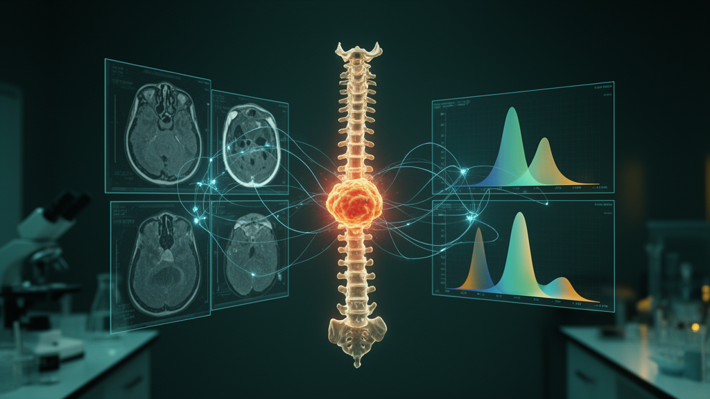
When breast cancer spreads to the spine, one of the most pressing questions a treating physician faces is deceptively simple: will chemotherapy actually work? The answer shapes everything that follows, from drug selection to radiation planning to the timing of surgery. Yet for decades, oncologists have had no reliable way to answer it before treatment begins. A new retrospective study from Peking University Cancer Hospital & Institute, published in BMC Medical Imaging, reports that a deep-learning model trained on pretreatment MRI data can predict chemotherapy response in spinal metastases with an accuracy that substantially outperforms the conventional diffusion parameters radiologists currently rely on.
The research, led by Hai-Tao Zhu, Yan-Jie Shi, Xiao-Ting Li and Ying-Shi Sun, enrolled 37 patients with breast cancer who harbored a total of 191 spinal metastatic lesions. Every patient underwent diffusion weighted imaging, or DWI, before chemotherapy started. DWI is a magnetic resonance technique that measures how freely water molecules move within tissue. In tumors, dense cell packing restricts water motion, and this restriction can be quantified. The team then split the patients into an investigating group of 19 and an independent test group of 18, a design that allowed them to build the model on one cohort and validate it on data the model had never seen.
The technical innovation at the heart of the study lies in how the imaging data were processed. Traditional DWI analysis acquires images at multiple b-values, which are gradient-strength settings that determine how sensitive each image is to water diffusion. Researchers typically fit the signal decay across these b-values into a predefined mathematical model, such as the apparent diffusion coefficient, the intravoxel incoherent motion model, the diffusion kurtosis model, or the stretched exponential model. Each model yields parameters like ADC, DDC, α, D*, D_true, frac, D_app and K_app, each intended to capture a different aspect of tissue microstructure. The problem is that these models impose assumptions on the data, and if the underlying biology does not match the assumed curve shape, information is lost or distorted.
Zhu and colleagues took a different route. Instead of fitting the multi-b-value signals into any model, they converted the raw signal intensities within each lesion into histograms, one histogram per b-value, and then concatenated all ten histograms into a single composite descriptor they call a histogram signature. This signature preserves the full distribution of signal intensities across the entire b-value range without forcing the data into a curve-fitting framework. Two radiologists manually delineated the borders of each lesion on the pretreatment DWI images to define the regions of interest, ensuring that the signatures were derived from tumor tissue rather than surrounding bone marrow or muscle.
The histogram signatures were then fed into a convolutional neural network, a class of deep-learning architecture that excels at detecting spatial and statistical patterns in structured data. The network’s task was binary classification: given a pretreatment signature, predict whether the corresponding patient’s disease would progress under chemotherapy, designated progressive disease or PD, or fall into the non-PD category. The network was trained on the investigating group and evaluated on the test group using receiver operating characteristic analysis, the standard method for quantifying diagnostic discrimination.
The results were striking. The histogram-signature model achieved an area under the ROC curve, or AUC, of 0.882, with a 95 percent confidence interval spanning 0.803 to 0.938. An AUC of 0.882 indicates strong discriminatory power, meaning the model could correctly rank a randomly chosen progressive-disease patient above a randomly chosen responder roughly 88 percent of the time. By comparison, the best-performing individual fitted parameter, D_app, reached an AUC of only 0.685, with a confidence interval of 0.585 to 0.774. The difference between the two approaches was statistically significant, with a Z statistic of 3.312 and a P value of 0.0009, confirming that the advantage of the histogram signature over the conventional parameter was not a chance finding.
Why does the model-free approach outperform the fitted parameters? The answer likely lies in what each method captures. A single fitted parameter like ADC or D_app compresses the entire diffusion signal decay into one number per voxel, discarding information about the shape of the decay curve, the heterogeneity within the tumor, and the distribution of signal values across the lesion. Histogram signatures, by contrast, retain the full statistical profile at every b-value. Tumors are notoriously heterogeneous, containing regions of necrosis, densely packed viable cells, and variable vascularity, all of which influence diffusion behavior in ways that a single scalar parameter cannot fully represent. By preserving this complexity, the signature gives the neural network a richer substrate from which to learn the patterns that distinguish responders from non-responders.
The clinical implications are considerable. Spinal metastases from breast cancer are common and often painful, and progressive disease in the spine can lead to vertebral collapse, spinal cord compression, and severe neurological impairment. If oncologists could identify, before the first cycle of chemotherapy, which patients are unlikely to respond, they could escalate to alternative regimens, add local therapies such as radiotherapy or ablation, or intensify surveillance for skeletal complications. Conversely, patients predicted to respond could proceed with confidence, avoiding unnecessary changes to a working plan. The pretreatment nature of the prediction is key: it moves decision-making from a reactive posture, waiting weeks for follow-up imaging to reveal whether the tumor is shrinking, to a proactive one grounded in data available on day one.
The study is not without limitations. Thirty-seven patients and 191 lesions constitute a modest sample size, and the retrospective design means the model was built and tested on historical data rather than prospectively enrolled patients. The radiologists’ manual delineation of lesion borders, while carefully performed, introduces a degree of operator dependence that automated segmentation could eventually reduce. External validation at other institutions, with different MRI scanners and imaging protocols, will be essential before the approach can be considered ready for routine clinical deployment. The authors note that the work was supported by the National Key Research and Development Program of China under grant 2023YFC3402805, and the study received ethics approval from Peking University Cancer Hospital with informed consent obtained from all enrolled subjects.
Nevertheless, the findings point toward a broader shift in oncological imaging. Rather than asking radiologists to extract a handful of predefined parameters and interpret them through clinical experience, the field is moving toward models that learn directly from the full richness of the imaging data. The histogram-signature approach demonstrated here sidesteps the long-standing tension between mathematical elegance and biological reality, showing that sometimes the best way to model diffusion is not to model it at all. If future prospective studies confirm these results, the combination of multi-b-value DWI and deep learning could become a standard pretreatment tool for patients facing spinal metastases, offering a data-driven answer to the question that matters most: will this chemotherapy work for me?
Subject of Research: Pretreatment prediction of the response to chemotherapy for spine metastases of breast cancer using histogram signatures from diffusion weighted imaging with multiple b-values: a retrospective study
Article Title: Pretreatment prediction of the response to chemotherapy for spine metastases of breast cancer using histogram signatures from diffusion weighted imaging with multiple b-values: a retrospective study
Article References: Zhu, H.-T., Shi, Y.-J., Li, X.-T., & Sun, Y.-S. (2026). Pretreatment prediction of the response to chemotherapy for spine metastases of breast cancer using histogram signatures from diffusion weighted imaging with multiple b-values: a retrospective study. BMC Medical Imaging. https://doi.org/10.1186/s12880-026-02767-5
Image Credits: AI Generated
DOI: 10.1186/s12880-026-02767-5
Keywords: Pretreatment, prediction, response, chemotherapy, spine, metastases, breast, cancer, histogram, signatures, diffusion, weighted
Cite Scienmag News
Nathaniel Bowman. (October 19, 2026). Deep Learning Reads MRI Histograms to Predict Which Spine Tumors Will Resist Chemotherapy. Scienmag. https://scienmag.com/deep-learning-reads-mri-histograms-to-predict-which-spine-tumors-will-resist-chemotherapy/
Nathaniel Bowman. "Deep Learning Reads MRI Histograms to Predict Which Spine Tumors Will Resist Chemotherapy." Scienmag, 19 October 2026, https://scienmag.com/deep-learning-reads-mri-histograms-to-predict-which-spine-tumors-will-resist-chemotherapy/. Accessed 19 October 2026.
Nathaniel Bowman. "Deep Learning Reads MRI Histograms to Predict Which Spine Tumors Will Resist Chemotherapy." Scienmag. October 19, 2026. https://scienmag.com/deep-learning-reads-mri-histograms-to-predict-which-spine-tumors-will-resist-chemotherapy/


