


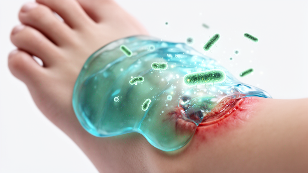
Diabetic foot ulcers represent a severe and often debilitating complication of diabetes mellitus, affecting a significant portion of the global diabetic population. These chronic wounds are characterized by delayed healing, persistent infection, and a high risk of lower-extremity amputation. Traditional treatment methods often struggle to maintain effective antibiotic concentrations at the wound site for the extended periods required for full recovery. A new study published in Pharmacia introduces a sophisticated thermoresponsive injectable gel designed to address these limitations by combining sustained antibiotic delivery with tissue-regenerative properties.
The research team developed a novel formulation that encapsulates levofloxacin hemihydrate within poly(lactic-co-glycolic acid) nanoparticles. These nanoparticles are then embedded in a thermoresponsive in situ gel matrix composed of Poloxamer 407 and Poloxamer 188. The system is engineered to remain a liquid at room temperature, allowing for easy syringeability and application to irregular wound surfaces. Upon contact with the body, the gel undergoes a sol-gel transition, forming a stable depot that retains the drug at the site of injury.
Optimization of the nanoparticle formulation was critical to achieving the desired therapeutic profile. The researchers evaluated twenty-five different formulations to identify the optimal balance of particle size, charge, and drug encapsulation. The selected formulation, designated as F21, exhibited a particle size of 77.1 nanometers and a zeta potential of +55.90 millivolts. This positive surface charge is particularly advantageous for wound applications, as it facilitates electrostatic interactions with negatively charged bacterial membranes and wound tissues, thereby enhancing local retention and antibacterial efficacy.
The encapsulation efficiency of the optimized nanoparticles reached 98.6 percent, ensuring that the vast majority of the levofloxacin was successfully loaded into the polymeric matrix. This high efficiency is crucial for maintaining therapeutic drug levels over time. The nanoparticles were characterized using dynamic light scattering and field-emission scanning electron microscopy, which confirmed their uniform nanoscale distribution and smooth morphology. Fourier-transform infrared spectroscopy further verified that the drug remained chemically stable within the polymer, with no significant destructive interactions observed.
Incorporating the nanoparticles into the thermoresponsive gel resulted in a system with favorable rheological properties. The gelation temperature ranged from 32.1 to 34.6 degrees Celsius, closely matching physiological skin temperatures. This ensures that the gel solidifies rapidly upon application, forming a protective barrier that conforms to the wound bed. The viscosity of the gel increased significantly at body temperature, reaching values up to 10,280 centipoise, which helps to prevent the premature leakage of the drug and ensures prolonged contact with the infected tissue.
In vitro release studies demonstrated that the formulation provided sustained delivery of levofloxacin over an extended period. While free drug solutions released the antibiotic rapidly, the nanoparticle-loaded gels maintained therapeutic concentrations for up to 240 hours. The release kinetics followed the Korsmeyer-Peppas model, indicating a combination of diffusion and polymer erosion mechanisms. This sustained release profile is particularly beneficial for diabetic ulcers, where maintaining antibiotic levels above the minimum inhibitory concentration for several days is essential to combat resistant bacterial strains and biofilms.
The study also investigated the impact of adding hyaluronic acid to the gel matrix. Hyaluronic acid is a naturally occurring glycosaminoglycan that plays a vital role in tissue hydration and cell migration. The formulation containing hyaluronic acid, designated as Formula A+, showed comparable drug release kinetics to the version without it. However, in vivo studies in diabetic mice revealed that Formula A+ produced the most significant therapeutic effects. The addition of hyaluronic acid enhanced tissue regeneration, promoted complete re-epithelialization, and reduced inflammatory cell infiltration, leading to superior wound closure compared to other groups.
Histopathological analysis of the treated wounds provided detailed insights into the healing process. Mice treated with Formula A+ exhibited near-complete restoration of skin architecture, with well-organized collagen deposition and minimal scar formation. In contrast, untreated diabetic wounds showed significant tissue damage, hyperkeratosis, and fibrosis. The improved healing outcomes were attributed to the synergistic effects of sustained antibiotic delivery and the biological activity of hyaluronic acid, which modulates inflammation and stimulates angiogenesis. These findings suggest that the combination of targeted drug delivery and tissue-regenerative agents can effectively address the complex pathophysiology of diabetic ulcers.
Despite the promising results, the researchers acknowledged several limitations that require further investigation. In vitro studies do not fully replicate the complex environment of a human diabetic wound, and the in vivo model used small rodents, which may not accurately represent human clinical outcomes. Additionally, long-term safety, systemic absorption, and repeated-dose toxicity need to be evaluated before the formulation can be considered for clinical translation. Scalable manufacturing processes and regulatory approval for combination nanomedicine wound dressings also present significant challenges that must be overcome.
This study highlights the potential of advanced drug delivery systems to improve the management of chronic diabetic wounds. By combining the benefits of sustained antibiotic release with the regenerative properties of hyaluronic acid, the developed gel offers a comprehensive approach to treating infected ulcers. Future research should focus on validating these findings in larger animal models and clinical trials, as well as assessing the formulation’s efficacy against bacterial biofilms. If successful, this technology could reduce the frequency of dressing changes, improve patient adherence, and ultimately lower the risk of amputation for patients suffering from diabetic foot ulcers.
Subject of Research: Thermoresponsive in situ gel for diabetic wound healing
Article Title: Hyaluronic acid-functionalized thermoresponsive in situ gel embedded with levofloxacin-loaded PLGA nanoparticles for enhanced diabetic wound healing
Article References: Mahmood Shihab, B., & Qasim Almajidi, Y. (2026). Hyaluronic acid-functionalized thermoresponsive in situ gel embedded with levofloxacin-loaded PLGA nanoparticles for enhanced diabetic wound healing. Pharmacia, 73, Article e205720. https://doi.org/10.3897/pharmacia.73.e205720
Image Credits: AI Generated
DOI: 10.3897/pharmacia.73.e205720
Keywords: diabetic ulcers, levofloxacin, PLGA nanoparticles, hyaluronic acid, drug delivery, wound healing, thermoresponsive gel, antibiotic therapy, Hyaluronic, acid-functionalized, thermoresponsive, situ
Cite Scienmag News
Ophelia Keating. (October 18, 2026). Smart Gel Accelerates Healing of Infected Diabetic Wounds. Scienmag. https://scienmag.com/smart-gel-accelerates-healing-of-infected-diabetic-wounds/
Ophelia Keating. "Smart Gel Accelerates Healing of Infected Diabetic Wounds." Scienmag, 18 October 2026, https://scienmag.com/smart-gel-accelerates-healing-of-infected-diabetic-wounds/. Accessed 18 October 2026.
Ophelia Keating. "Smart Gel Accelerates Healing of Infected Diabetic Wounds." Scienmag. October 18, 2026. https://scienmag.com/smart-gel-accelerates-healing-of-infected-diabetic-wounds/


