


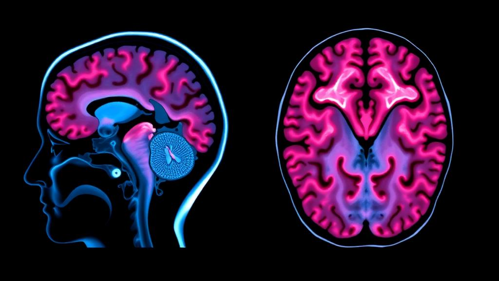
Duchenne and Becker muscular dystrophies have always been described as diseases of muscle, yet a growing body of evidence shows that the same genetic defect quietly reshapes the brain. A new review published in the Journal of Neurology by Marion Lancien, Cyrille Vaillend and colleagues from the University of Montpellier, CHU de Montpellier and Université Paris-Saclay brings together decades of brain imaging results from patients with dystrophinopathies and from animal models, offering the most complete picture to date of how the loss of dystrophin proteins alters brain structure, function and connectivity. The work arrives at a moment when gene therapies targeting the DMD gene are moving rapidly through clinical trials, making it more urgent than ever to understand exactly what happens inside the brains of affected individuals.
The DMD gene is the largest known protein-coding gene in the human genome, and it does not produce a single protein but a family of them. In muscle, the full-length dystrophin Dp427 anchors the dystrophin-glycoprotein complex, a transmembrane linker between laminin and actin that stabilizes muscle fibers during contraction. In the brain, however, the picture is far more complex. Several dystrophin isoforms are expressed in neural tissue, including Dp427 itself, the shorter Dp140, and the tiny Dp71, each with its own developmental timing and cellular localization. Dp427 appears in neurons, particularly in the postsynaptic density of excitatory synapses in the hippocampus and cortex, where it helps organize receptor clusters. Dp140 is expressed in developing fetal brain and in glial cells, while Dp71 is found in astrocytes and other macroglial cells, where it anchors aquaporin-4 channels and potassium channels critical for water and ion homeostasis. Mutations in the DMD gene can therefore disrupt one or several of these brain dystrophins depending on where the genetic lesion falls, producing a spectrum of cognitive and neuropsychiatric comorbidities that range from mild learning difficulties to intellectual disability, autism spectrum features, attention deficit hyperactivity disorder and anxiety.
The review synthesizes imaging findings across magnetic resonance imaging, diffusion tensor imaging, positron emission tomography, single-photon emission computed tomography and magnetic resonance spectroscopy, applied both to patients and to animal models including mdx mice, DMD rats and DE50-MD dogs. One of the most consistent findings is the presence of mild cortical atrophy, particularly in frontal regions, together with slight modifications in the volumes of specific brain structures. Studies of boys with Duchenne muscular dystrophy have shown reduced cerebral gray matter and altered white matter compared with typically developing controls, and voxel-based morphometric analyses in Becker muscular dystrophy patients reveal similar though generally milder structural changes. In the motor cortex specifically, decreased gray matter concentration and reduced local synchronization of spontaneous neural activity have been documented, suggesting that the brain regions controlling movement are affected in parallel with the muscles they command.
Beyond gross anatomy, the imaging evidence points to profound alterations in brain physiology. Cerebral blood flow is decreased in patients with Duchenne muscular dystrophy, with frontal lobe-dominant reductions that appear to be progressive over time according to longitudinal observations in Japanese patient cohorts. Brain hypometabolism has been detected through positron emission tomography studies showing altered regional glucose metabolism, and magnetic resonance spectroscopy has revealed abnormalities in brain biochemistry, including changes in phosphorus-31 and proton spectra that suggest disturbed energy metabolism and neuronal integrity. Animal models reinforce these observations: mdx mice show abnormal cerebral diffusion and perfusion in vivo, and the DMD rat exhibits measurable disruptions in both microstructure and neurochemistry. The review also highlights an abnormal cerebral response to stress, with recent work demonstrating that brain glucose metabolism serves as a neuronal substrate of the abnormal behavioral response to stress in the mdx mouse, providing a biological explanation for the heightened anxiety and stress reactivity frequently reported in patients.
White matter microstructure alterations emerge as another central theme. Diffusion tensor imaging studies in children with Duchenne muscular dystrophy consistently show abnormal fractional anisotropy and diffusivity in major white matter tracts, indicating disrupted myelination or axonal organization. This aligns with cellular evidence from mdx mice, where myelination is delayed during postnatal brain development and oligodendrogenesis is defective, with persistent hypomyelination observed even in adult animals. Advanced diffusion approaches probing the diffusion of water and metabolites have further refined the assessment of white matter microstructure in patients, suggesting that the alterations reflect genuine tissue-level changes rather than artifacts of image processing. The review notes that these white matter changes may underlie some of the cognitive profiles seen in patients, particularly difficulties with executive function and processing speed that depend on efficient long-range communication between brain regions.
Functional connectivity disturbances add yet another dimension to the imaging picture. Resting-state functional MRI has revealed altered default-mode network functional connectivity in Duchenne muscular dystrophy patients, and connectomic analyses in patients with mild cognitive impairment show broader large-scale network disturbances. Interestingly, the review draws parallels with connectivity patterns observed in other neurodevelopmental and psychiatric conditions, including attention deficit hyperactivity disorder, obsessive-compulsive disorder and autism, where both hypoconnectivity and hyperconnectivity of specific networks have been documented. This suggests that the loss of brain dystrophins may produce network-level imbalances that resemble, at least partially, the connectomic signatures of common neuropsychiatric disorders, providing a mechanistic bridge between the genetic lesion and the psychological symptoms that patients experience.
A crucial insight from the review concerns the genotype-phenotype relationship, particularly the role of Dp140. When mutations affect Dp427 alone, cognitive and neuropsychiatric consequences tend to be milder, but when Dp140 expression is also disrupted, severity increases substantially. This is consistent with the developmental timing of Dp140, which is expressed during fetal brain development and appears to play a critical role in glutamatergic transmission and social behaviour, as demonstrated in the mdx52 mouse model that lacks Dp140. Studies comparing patients with mutations at different locations along the DMD gene have confirmed that the risk of cognitive impairment correlates with mutation location, and that Dp71 disruption contributes to the severity of intellectual disability. The review emphasizes that the variable severity of cerebral disorders depends on which combination of brain dystrophins is lost, making the DMD gene a remarkable example of a single gene producing multiple phenotypes through its family of protein products.
The question of progression remains one of the most clinically important aspects of the review. Some correlations between imaging parameters and cognitive or psychological impairments have been reported, but the authors caution that these remain to be confirmed in larger cohorts. Evidence for progression comes from longitudinal observations showing that frontal lobe-dominant cerebral blood flow reduction and atrophy can worsen over time in Duchenne muscular dystrophy, raising the possibility that cerebral involvement is not static but evolves alongside the muscular disease. However, other spectroscopy studies indicate preserved cerebral biochemical composition in some patient groups, suggesting that the trajectory may be more nuanced than simple deterioration. The review calls for standardized longitudinal imaging protocols that would allow researchers to track cerebral changes across the lifespan of patients, from early childhood through adulthood, and to determine whether therapeutic interventions targeting the muscle disease also benefit the brain.
The implications of this work extend well beyond academic interest. As exon-skipping therapies, gene replacement strategies and gene-editing approaches advance through clinical trials for Duchenne muscular dystrophy, the question of whether these treatments restore brain dystrophins becomes paramount. Many current therapies are designed primarily with muscle in mind, and the blood-brain barrier poses a significant challenge for delivering dystrophin-restoring treatments to the central nervous system. The imaging biomarkers catalogued in this review, from cortical atrophy to cerebral blood flow, white matter microstructure, brain metabolism and functional connectivity, provide a toolkit for evaluating whether future therapies reach the brain and produce measurable benefits. Without such biomarkers, it would be impossible to know whether a treatment that improves muscle strength also improves learning, memory or emotional regulation.
The review ultimately reframes dystrophinopathies as genuine brain diseases with muscular consequences rather than the reverse. The convergence of structural, physiological, metabolic and connectomic imaging evidence, replicated across species and imaging modalities, establishes that the loss of brain dystrophins produces real and measurable cerebral alterations. While the correlations between imaging findings and cognitive or psychological symptoms await confirmation in larger studies, the biological plausibility is strong, supported by cellular and molecular evidence of dystrophin function in neurons, glia and the neurovascular unit. As the authors conclude, cerebral imaging has shed new light on the impact of dystrophinopathies on brain structure, function and connectivity, and this light now guides the search for therapies that address the full spectrum of what it means to live with a dystrophin mutation.
Subject of Research: Neuroscience
Article Title: Brain imaging in dystrophinopathies
Article References: Lancien, M., Leboucq, N., Desguerre, I., Rivier, F., & Vaillend, C. (2026). Brain imaging in dystrophinopathies. Journal of Neurology, 273(11), Article 669. https://doi.org/10.1007/s00415-026-14215-z
Image Credits: AI Generated
DOI: 10.1007/s00415-026-14215-z
Keywords: Duchenne muscular dystrophy, Becker muscular dystrophy, brain imaging, dystrophin, MRI, cognitive impairment, Brain, imaging, dystrophinopathies, scientific research
Cite Scienmag News
Cassandra Pierce. (October 17, 2026). Brain imaging in dystrophinopathies. Scienmag. https://scienmag.com/brain-imaging-in-dystrophinopathies/
Cassandra Pierce. "Brain imaging in dystrophinopathies." Scienmag, 17 October 2026, https://scienmag.com/brain-imaging-in-dystrophinopathies/. Accessed 17 October 2026.
Cassandra Pierce. "Brain imaging in dystrophinopathies." Scienmag. October 17, 2026. https://scienmag.com/brain-imaging-in-dystrophinopathies/


