


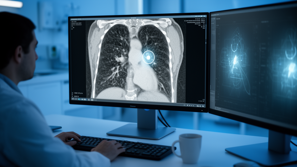
Lung cancer remains one of the deadliest malignancies worldwide, and much of its lethality stems from a deceptively simple problem: seeing the tumor clearly. On computed tomography scans, lung tumors often blur into surrounding tissue, their edges fading into inflammation, scar tissue, or healthy lung parenchyma. A new study published in Scientific Reports by M. Sutharsan of Dhirajlal Gandhi College of Technology and K. R. Kavitha of Sona College of Technology, both in Salem, Tamil Nadu, India, tackles this problem head-on with an integrated artificial intelligence framework called BAMR-HF, which combines boundary-aware segmentation, hybrid radiomic–deep feature fusion, and context-aware classification into a single pipeline for reading lung CT images.
The framework is built from three cooperating neural networks, each addressing a different failure mode of automated tumor analysis. The first, BAMR-Net, performs the segmentation task: it draws the precise outline of the tumor within a CT slice. Rather than treating this as a generic pixel-labeling problem, the network is designed to be boundary-aware, meaning it explicitly attends to the transition zone between tumor and healthy tissue. It uses multi-scale residual learning, a technique in which the network processes the image at several spatial resolutions simultaneously and uses residual connections to preserve fine detail as information flows through increasingly abstract layers. This matters because tumors can appear as small nodules or large masses, and a network that only looks at one scale will miss features visible at another.
Boundary refinement is the second pillar of BAMR-Net. In medical image segmentation, the difference between a clinically useful contour and a dangerous one often comes down to a few pixels at the tumor margin. Conventional convolutional networks tend to produce blurry edges because their downsampling steps discard spatial precision. By incorporating a dedicated boundary-refinement stage, the architecture sharpens the predicted contour after the coarse segmentation is produced, recovering the crisp demarcation that radiologists rely on when judging whether a nodule is invading adjacent structures.
Once the tumor is segmented, the framework hands the region to TRFF-Net, which performs classification by fusing two fundamentally different kinds of features. The first kind is radiomic: mathematically defined descriptors such as texture statistics, shape measures, and intensity distributions that radiomics researchers have long used to quantify tumor appearance. The second kind is deep: the abstract, learned representations that a convolutional network discovers on its own during training. Neither alone tells the full story. Handcrafted radiomic features are interpretable and stable but limited to what engineers thought to measure; deep features are expressive but opaque and sometimes brittle. TRFF-Net combines them through learnable weighted fusion, meaning the network itself learns during training how much to trust each feature family for each decision, rather than a human fixing the weights in advance.
The third component, MTCA-Net, adds something radiologists do instinctively but most automated systems ignore: context. A tumor does not exist in isolation. The tissue immediately surrounding it—the peritumoral region—carries diagnostic clues, from the texture of adjacent lung to signs of invasion or inflammation. MTCA-Net incorporates both tumor and peritumoral contextual information into its classification decision, effectively giving the network a wider field of view than the segmented lesion alone. This context-aware design reflects a growing recognition in computational pathology that the microenvironment around a lesion can be as informative as the lesion itself.
The researchers evaluated the complete BAMR-HF framework on a processed Lung Cancer Segmentation Dataset with Lung-RADS classes comprising 972 CT images. The data were split into a training partition of 708 images and a repository-defined held-out test partition of 264 images, meaning the final numbers were computed on data the networks never saw during training. The source labels, which follow the Lung-RADS reporting system used in lung cancer screening, were consolidated into three severity categories: Mild (LR2), Moderate (LR3), and Severe (LR4A–LR4B). This consolidation turns a fine-grained radiological grading scheme into a tractable classification problem while preserving the clinically meaningful distinction between low- and high-concern findings.
The results are striking. BAMR-Net achieved a test Dice score of 0.8563, a standard overlap metric where 1.0 represents perfect agreement between the predicted tumor contour and the ground-truth annotation. On the classification side, TRFF-Net and MTCA-Net achieved test accuracies of 0.9038 and 0.9327 respectively, showing that the context-aware network outperformed the fusion network alone. The complete BAMR-HF framework, combining all three stages, attained an accuracy of 0.9478, an F1-score of 0.9476, an area under the receiver operating characteristic curve (AUC) of 0.9872, and a Matthews correlation coefficient (MCC) of 0.9215. The MCC is particularly notable because it is robust to class imbalance and near 1.0 indicates near-perfect agreement between prediction and truth across all categories.
These numbers suggest that the three design choices—boundary awareness, hybrid feature representation, and contextual modeling—are genuinely complementary rather than redundant. Each network contributes information the others lack: the segmentation network defines what to analyze, the fusion network determines how to characterize it, and the context network situates it in its surroundings. The progression from TRFF-Net’s 0.9038 accuracy to the full framework’s 0.9478 indicates that the added stages carry real diagnostic weight, not just architectural complexity.
The authors are careful about the limits of their claims, and rightly so. The study was conducted on a single processed dataset, and the abstract explicitly notes that broader validation on independent multi-institutional data remains necessary before the framework can be considered ready for clinical deployment. This caveat is standard and important: deep learning systems in medical imaging frequently perform well on curated datasets but degrade when confronted with scans from different scanners, protocols, or patient populations. The published version is also being shared early as an accepted manuscript, subject to further edits before the final Version of Record appears.
Even with those caveats, the study offers a compelling template for how automated lung tumor analysis might evolve. Rather than betting everything on ever-larger deep networks, BAMR-HF demonstrates the value of architectural humility: explicitly encoding what radiologists know about boundaries, texture, and context, then letting learnable fusion decide how to weigh that knowledge against what the network discovers on its own. As lung cancer screening programs expand and radiologists face growing reading volumes, frameworks that combine handcrafted domain knowledge with learned representations—and that are honest about the validation still required—may prove to be exactly the kind of tool the clinic needs.
Subject of Research: Health
Article Title: Boundary-aware multi-scale residual learning with hybrid radiomic–deep fusion for lung tumor segmentation and classification
Article References: Sutharsan, M., & Kavitha, K. R. (2026). Boundary-aware multi-scale residual learning with hybrid radiomic–deep fusion for lung tumor segmentation and classification. Scientific Reports. https://doi.org/10.1038/s41598-026-74349-y
Image Credits: AI Generated
DOI: 10.1038/s41598-026-74349-y
Keywords: lung cancer, medical imaging, deep learning, radiomics, CT segmentation, artificial intelligence, tumor classification, BAMR-HF, Boundary-aware, multi-scale, residual, learning
Cite Scienmag News
Nathaniel Bowman. (October 17, 2026). AI Framework Reads Lung Tumors With Unusual Precision on CT Scans. Scienmag. https://scienmag.com/ai-framework-reads-lung-tumors-with-unusual-precision-on-ct-scans/
Nathaniel Bowman. "AI Framework Reads Lung Tumors With Unusual Precision on CT Scans." Scienmag, 17 October 2026, https://scienmag.com/ai-framework-reads-lung-tumors-with-unusual-precision-on-ct-scans/. Accessed 17 October 2026.
Nathaniel Bowman. "AI Framework Reads Lung Tumors With Unusual Precision on CT Scans." Scienmag. October 17, 2026. https://scienmag.com/ai-framework-reads-lung-tumors-with-unusual-precision-on-ct-scans/


