


A 10-year-old female spayed mixed-breed dog walked into a veterinary neurology service with seizures, stumbling, and a week of strange rigid episodes — and within 24 hours of diagnosis was dead from a brain hemorrhage so rare that no comparable case had ever been documented in the veterinary literature. Clinicians at Cornell University’s College of Veterinary Medicine report in Frontiers in Veterinary Science that the dog’s fatal intracranial subdural hemorrhage was secondary to primary immune-mediated thrombocytopenia, or ITP, a disorder in which the immune system destroys the body’s own platelets. The case is the first in veterinary medicine to document severe, fatal cerebral subdural hemorrhage as a primary neurological manifestation of canine ITP, and it carries a blunt warning for practitioners: an acute, progressive neurological presentation should prompt a platelet count check until ITP is ruled out.
ITP is one of the most common hemostatic disorders seen in dogs. In its primary form, the immune system mistakenly tags platelets for destruction, and in its secondary form another underlying disease or drug triggers the same loss. Spontaneous hemorrhage typically does not occur until the platelet count falls below roughly 30,000 per microliter, and the clinical picture is notoriously variable: lethargy, inappetence, fever, and the classic pinpoint petechiae and larger ecchymoses of the skin and mucous membranes are well documented, while less frequent signs include melena, blood in the urine, nosebleeds, and bleeding into the eye. Studies of canine ITP generally report a fair prognosis for survival to discharge, but morbidity is variable and mortality can reach 27 percent. Neurological signs from intracranial hemorrhage, however, are uncommon to exceptionally rare in both human and veterinary literature — which is precisely what makes this case remarkable.
The dog’s story began about a month before referral, when the owner noticed decreased activity, reluctance to jump, and intermittent front-limb lameness. The primary veterinarian, suspecting osteoarthritis, prescribed the non-steroidal anti-inflammatory drug meloxicam and recommended referral if things did not improve. Instead, over the following week the dog developed progressive seizure-like episodes: whole-body rigidity with non-responsive mentation lasting 15 to 20 minutes, occurring a few times daily. Blood work at an emergency clinic revealed a mild leukocytosis and severe thrombocytopenia — a platelet count of 21,000 per microliter against a reference interval of 170,000 to 400,000 — with a few small platelet clumps on the smear. The dog was negative on screening for heartworm antigen and for antibodies against Borrelia burgdorferi, Anaplasma, and Ehrlichia, the major vector-borne mimics of immune-mediated disease.
On transfer to the neurology service, the picture sharpened. The dog was dull, carried its head low, and showed ambulatory tetraparesis with vestibular ataxia, a tendency to turn left, a mild left head tilt, and positional nystagmus shifting between vertical and rotary forms. Cranial nerve examination revealed absent menace responses bilaterally with moderate to severe mydriasis, though pupillary light reflexes remained intact. Fundic examination showed bilateral retinal hemorrhage, worse in the right eye, and physical examination found two ecchymoses on the left thoracic region. The neuroanatomical localization was multifocal brain disease, involving the left forebrain and brainstem. Repeat hematological evaluation was striking: a platelet count of just 9,000 per microliter with no visible clumps and a markedly increased mean platelet volume of 26.8 femtoliters, a signature of young, large platelets being released by the marrow in a desperate compensatory effort. Reticulocytosis with a normal hematocrit confirmed the marrow was responding to ongoing platelet loss rather than failing.
The diagnostic workup then proceeded along the lines of the American College of Veterinary Internal Medicine consensus statement on ITP, which remains a diagnosis of exclusion. A comprehensive coagulation profile — activated partial thromboplastin time, prothrombin time, quantitative fibrinogen by the Clauss method, antithrombin activity, and quantitative D-dimers — was entirely within normal limits, ruling out concurrent consumption coagulopathies such as disseminated intravascular coagulation. Thoracic radiographs were unremarkable, and abdominal ultrasound showed only a moderately enlarged, heterogeneous spleen, interpreted as a benign reactive change such as lymphoid hyperplasia or extramedullary hematopoiesis. The timeline also effectively excluded meloxicam as the inciting trigger, since both the severe thrombocytopenia and the clinical signs were present before the drug was given. With no other cause identified, the team established a presumptive diagnosis of primary ITP.
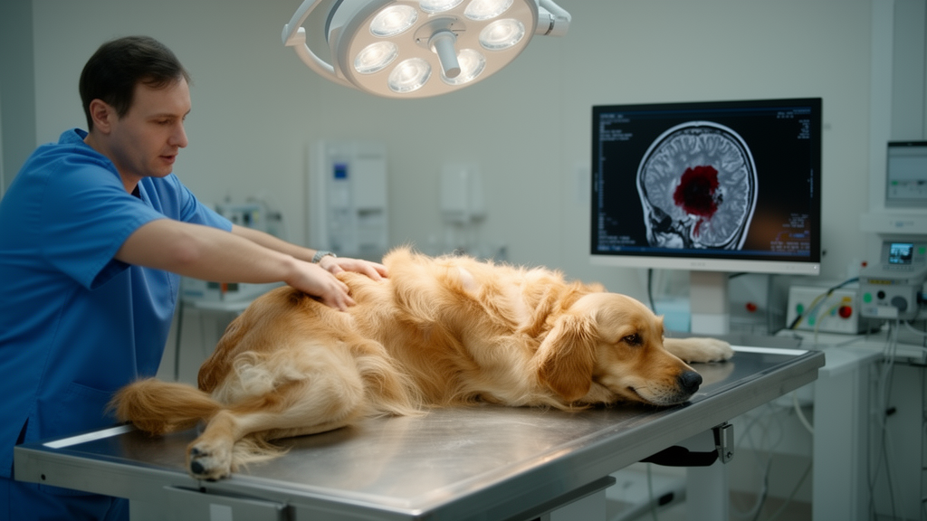
Brain MRI, performed the same day under general anesthesia on a 1.5-T scanner, delivered the case’s central finding. Minimally expanded, bilateral crescent-shaped, extra-axial fluid accumulations overlay the entire dorsolateral convexity of both cerebral hemispheres, not extending along the medial or basal borders. The collections were homogeneously hyperintense on T1-weighted, T2-weighted, and FLAIR images, with peripheral susceptibility artifact on T2*-weighted gradient-recalled echo sequences — a combination characteristic of late subacute intracranial subdural hemorrhage. The cerebral hemispheres were mildly compressed and diffusely swollen without midline shift, but the swollen cerebrum displaced the diencephalon and mesencephalon caudally, compressed the rostral cerebellum, and pushed the cerebellum toward, but not through, the foramen magnum. Imaging at the level of the tentorium confirmed that caudal displacement was restricted to the midline tectum, without full transtentorial herniation. The cervical spinal cord was mildly swollen with T2 hyperintensity, interpreted as secondary edema. Intracranial pressure was estimated to be high, and cerebrospinal fluid analysis was deliberately withheld given the severe thrombocytopenic state and suspected pressure elevation.
The MRI chronology is technically elegant and diagnostically crucial. On MRI, the signal intensity of intracranial hemorrhage is dictated primarily by the T1 relaxation and T2* susceptibility effects of hemoglobin degradation over time: oxyhemoglobin gives way to deoxyhemoglobin, then intracellular methemoglobin, extracellular methemoglobin, and eventually ferritin and hemosiderin, each stage altering the magnetic properties of the hematoma in predictable ways. The T1 and T2 hyperintensity with susceptibility artifact seen here placed the hemorrhage firmly in the late subacute stage, which spans days to months. Critically, the dog’s seizure episodes began approximately one week before imaging — a timeline that correlates closely with the late subacute to early chronic hemorrhage window and further validates severe thrombocytopenia and primary ITP as the etiology of the neurological signs, rather than a separate process.
Then came the devastating turn. Anesthesia and recovery were uneventful, and the dog was standing and walking on its own in the intensive care kennel with quiet mentation. A single dose of dexamethasone sodium phosphate had been given during recovery as the first immunomodulatory treatment. Three hours after recovery, on repeat neurological evaluation, the dog was acutely and severely obtunded with moderate respiratory effort, though cardiopulmonary auscultation was unremarkable. This was followed by abrupt bradypnea, cyanosis of the tongue, and respiratory arrest, rapidly progressing to cardiopulmonary arrest. Given the owner’s do-not-resuscitate status, CPR was not performed, and a postmortem examination was declined. In the absence of necropsy, a definitive cause of death could not be confirmed, though the authors consider ongoing hemorrhage or brain herniation — a rapid rise in intracranial pressure suppressing the medullary respiratory center — the most plausible mechanism. They also reason that pulmonary thromboembolism, a recognized differential for sudden death, typically presents with hyperventilation and tachypnea rather than the abrupt bradypnea observed here, making brainstem dysfunction the stronger explanation.
The case gains additional weight from comparison with the only two previously documented ITP-related hemorrhagic myelopathies in dogs. In one, a dog with a platelet count of 3,000 per microliter developed progressive tetraparesis; MRI revealed multifocal spinal cord hemorrhage, and despite resolution of the thrombocytopenia with immunomodulatory therapy, the neurological status deteriorated to tetraplegia and the dog was eventually euthanized. Postmortem examination confirmed hemorrhage throughout the entire spinal cord. In the second, a dog with acute tetraplegia was treated with dexamethasone and cyclosporine; the platelet count normalized by day five, and at five-week follow-up the dog showed only mild ataxia with a normal platelet count. Together, these cases illustrate the central paradox of ITP: there is no direct correlation between the severity of thrombocytopenia and the anatomical location or clinical severity of hemorrhage, and normalizing the platelet count does not guarantee neurological recovery. Notably, neither melena nor elevated blood urea nitrogen — the recognized poor-prognosis indicators in canine ITP — was present in the Cornell case, underscoring how little veterinary medicine currently knows about which ITP patients will develop catastrophic central nervous system bleeding.
In human medicine, intracranial hemorrhage in severe thrombocytopenic ITP patients carries an estimated mortality of 25 to 55 percent, and there is limited consensus on optimal management, particularly regarding emergent surgical decompression during rapid neurological deterioration; only a single human case report documents an excellent outcome following combined emergency splenectomy and craniotomy. No such surgical guidelines exist for veterinary patients, and the Cornell team notes that the rapid deterioration in this case — within 24 hours of diagnosis — left no realistic window for intervention. The authors’ conclusion is direct: although canine ITP generally carries a favorable prognosis when managed appropriately, life-threatening secondary intracranial hemorrhage can occur, and primary ITP should be considered as a differential diagnosis for any acute, progressive neurological presentation until a normal platelet count is confirmed. For a disease usually caught through skin and mucosal bleeding, this case is a stark reminder that the immune system’s attack on platelets can, rarely, reach the brain itself.
Subject of Research: Immune-mediated thrombocytopenia in veterinary medicine
Article Title: Case Report: Presumptive subdural hemorrhage as a rare neurological manifestation of primary immune-mediated thrombocytopenia in a dog
Article References: Hong, H.-P., Kei, T. G., Gagnon, N. A., & Scrivani, P. V. (2026). Case Report: Presumptive subdural hemorrhage as a rare neurological manifestation of primary immune-mediated thrombocytopenia in a dog. Frontiers in Veterinary Science, 13, Article 1969040. https://doi.org/10.3389/fvets.2026.1969040
Image Credits: AI Generated
DOI: 10.3389/fvets.2026.1969040
Keywords: Case, Report, Presumptive, subdural, hemorrhage, rare, neurological, manifestation, primary, immune-mediated, thrombocytopenia, scientific research
Cite Scienmag News
William Thompson. (October 17, 2026). Fatal Brain Bleed Revealed as Rare First-of-Its-Kind Complication of Canine Immune Disease. Scienmag. https://scienmag.com/fatal-brain-bleed-revealed-as-rare-first-of-its-kind-complication-of-canine-immune-disease/
William Thompson. "Fatal Brain Bleed Revealed as Rare First-of-Its-Kind Complication of Canine Immune Disease." Scienmag, 17 October 2026, https://scienmag.com/fatal-brain-bleed-revealed-as-rare-first-of-its-kind-complication-of-canine-immune-disease/. Accessed 17 October 2026.
William Thompson. "Fatal Brain Bleed Revealed as Rare First-of-Its-Kind Complication of Canine Immune Disease." Scienmag. October 17, 2026. https://scienmag.com/fatal-brain-bleed-revealed-as-rare-first-of-its-kind-complication-of-canine-immune-disease/


