


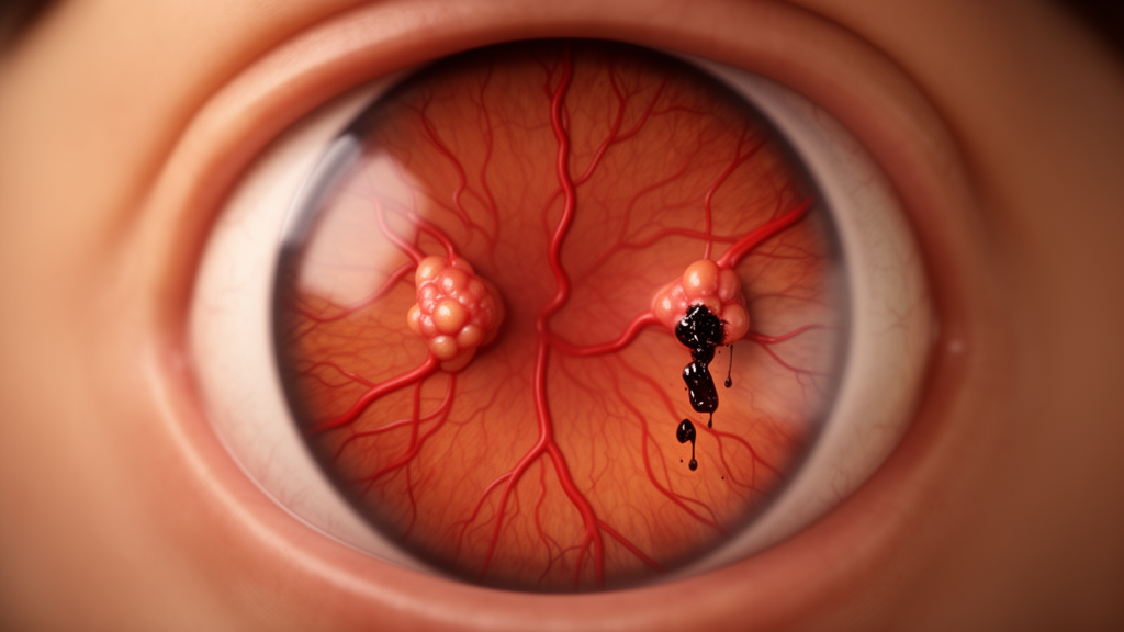
In a striking illustration of how unpredictable eye disease can be, ophthalmologists in Chongqing, China have documented a rare case in which two aneurysms on the retinal arteries of a single eye followed dramatically different courses over five years of follow-up. One lesion hardened into a fibrotic scar that permanently disturbed the retina’s architecture, while the other vanished completely, apparently in response to aggressive control of the patient’s blood pressure. The case, published in Frontiers in Medicine, offers one of the longest documented looks at the natural history of multiple retinal arterial macroaneurysms in one eye, and it carries a practical message: sometimes the best treatment is knowing when not to treat.
Retinal arterial macroaneurysm, or RAM, is an uncommon acquired disorder in which a major retinal artery develops a saccular or fusiform bulge, most often at the crossings of arteries and veins along the first to third order branches of the central retinal artery. First described by Robertson in 1973, the condition affects roughly one person in 9,000, with a strong predilection for older adults and women. More than 90 percent of patients are over 60, about 73 percent are women, and in roughly 90 percent of cases only one eye is involved. The pathogenesis is tightly linked to systemic cardiovascular disease, particularly hypertension, arteriosclerosis, and hyperlipidemia, with between 51 and 75 percent of affected patients carrying a diagnosis of high blood pressure.
The trouble with RAM is that it usually announces itself with a catastrophe. When the weakened arterial wall ruptures, blood can pour into multiple compartments of the eye simultaneously: beneath the retina, within its layers, in front of it, and into the vitreous cavity that fills the globe. Subretinal hemorrhage carries the worst prognosis, and animal studies have shown that photoreceptors begin to suffer significant damage within 24 hours of exposure, with irreversible injury to photoreceptors and the retinal pigment epithelium following if the blood remains for seven to fourteen days. Because the resulting hemorrhagic picture mimics so many other retinopathies, the misdiagnosis rate for RAM has been reported to reach 75 percent.
That is precisely what happened to the 65-year-old woman at the center of this report. She arrived with a one-day history of sudden floaters and a dark shadow in her left eye, and initially denied any history of hypertension, diabetes, or hyperlipidemia. Her best-corrected visual acuity was 0.8 in the right eye but only 0.25 in the left, and optical coherence tomography revealed an extensive hemorrhagic elevation of the retina in the inferotemporal macula. She was given oral iodized lecithin to promote blood resorption and sent for observation. Within weeks, however, the picture deteriorated rapidly: her vision fell to hand motions, then to mere light perception, and her blood pressure was recorded at a dangerous 187/104 mmHg, revealing previously undiagnosed and uncontrolled hypertension.
With dense vitreous hemorrhage obscuring any view of the fundus, the surgical team at a tertiary ophthalmic center performed a pars plana vitrectomy combined with internal limiting membrane peeling, intravitreal injection of recombinant tissue plasminogen activator to dissolve clot, fluid-air exchange, and silicone oil tamponade. The recovery was rocky. Recurrent bleeding extended from the vitreous cavity through the pupil into the anterior chamber, producing a hyphema that blocked the drainage angle and drove her intraocular pressure to 46 mmHg. Intravenous mannitol and oral acetazolamide failed to fully control the pressure, and surgeons ultimately performed anterior chamber irrigation to wash out the blood before corneal blood staining could set in.
It was only months later, once the ocular media had cleared enough for imaging, that the true diagnosis emerged. Combined fluorescein fundus angiography and indocyanine green angiography, the diagnostic gold standard for RAM, revealed two distinct hemorrhagic foci in the left eye. One lesion appeared as a well-demarcated hypofluorescent area with mild peripheral leakage in the late phase, while the second showed a faint hyperfluorescent outline without active leakage. The imaging team concluded that the mild peripheral leakage represented reactive staining from the abnormal retinal architecture caused by a large intraretinal blood clot, not the active vascular leakage that would demand laser treatment. On that basis, the clinicians made a deliberate decision to withhold laser photocoagulation and simply observe, a choice that the subsequent four years would vindicate.
What followed was a natural experiment unfolding inside a single eye. The initial lesion, which had been present at the time of the severe hemorrhage and before blood pressure was controlled, gradually evolved into an elevated fibrotic scar. Over more than four years of serial imaging, the height of the retinal elevation declined slowly, but the apex showed marked hyperreflectivity and persistent intraretinal cystoid spaces remained around the lesion. The second, de novo lesion, detected relatively early with only limited hemorrhage and managed under strict antihypertensive therapy with nifedipine and metoprolol, was no longer identifiable on optical coherence tomography by the eighth visit in October 2021. The surrounding retina remained flat, and the lesion had effectively resolved without any direct intervention.
The authors are careful about what this contrast does and does not prove. The two lesions differed not only in blood pressure exposure but also in hemorrhage severity, timing of detection, treatment history, and probably disease stage, and spontaneous regression of RAM is well recognized in the literature. Most patients with RAM experience visual improvement even without intervention, and some patients with concurrent hypertension have achieved complete recovery through antihypertensive therapy alone. Blood pressure control, the authors conclude, should be regarded as an important possible contributing factor to the divergent outcomes rather than a decisive prognostic determinant in this single case. Still, the pattern of persistence versus resolution is compelling, and it underscores the potential importance of early systemic blood pressure management in shaping the long-term prognosis of this retinal disease.
The case also carries a first: the earliest reported occurrence of nonvascular cystoid maculopathy secondary to suspected RAM. This rare entity involves intraretinal cystic spaces that form not from leaking blood vessels but from pathological factors that compromise the adhesion between retinal cells or impair the ability of the retinal pigment epithelium and Müller cells to keep the retina dehydrated. Fluorescein angiography is essential for telling it apart from ordinary cystoid macular edema caused by blood-retinal barrier disruption, because fluorescein does not leak into the cystic spaces of the nonvascular form. The condition is usually associated with non-exudative age-related macular degeneration, macular telangiectasia type 2, hereditary retinal dystrophies such as X-linked retinoschisis, optic nerve disorders, drug toxicities from agents like tamoxifen, and vitreomacular traction disorders. Seeing it arise next to a macroaneurysm is unprecedented, and the authors propose that tractional forces from the cicatricial lesion on the surrounding retina may have compromised Müller cell function, though they acknowledge the mechanism remains hypothetical without histopathological confirmation.
For clinicians, the practical lesson is about restraint guided by imaging. Laser photocoagulation, the mainstay of RAM treatment, promotes intra-aneurysmal thrombosis and vessel wall fibrosis, but it resolves macular edema slowly and carries risks including aneurysm rupture, distal vascular occlusion, and visual field defects, and some studies have even reported visual deterioration after treatment. Anti-VEGF injections, which this patient declined, are widely used but not always necessary. In this case, multimodal imaging showing two stable, non-leaking lesions allowed the team to avoid overtreatment entirely, and the patient’s visual acuity and lesion morphology remained stable through more than four years of observation. The authors also candidly list the limitations of a single retrospective case: no matched control group, incomplete early angiographic evidence, a non-standardized treatment regimen, and the possibility that regression reflected natural involution rather than any therapeutic effect. Even so, the five-year record stands as a valuable real-world window into the heterogeneous behavior of retinal arterial macroaneurysms, and a reminder that in the delicate plumbing of the human retina, timing and blood pressure may matter as much as any scalpel or laser.
Subject of Research: Divergent five-year outcomes of two retinal arterial macroaneurysms in a single eye with secondary nonvascular cystoid maculopathy
Article Title: Two suspected sequential retinal arterial macroaneurysms in a single eye with secondary nonvascular cystoid macular edema: 5-year follow-up: a case report
Article References: Chi, K., Lei, D., Guo, T., Shui, X., & Li, H. (2026). Two suspected sequential retinal arterial macroaneurysms in a single eye with secondary nonvascular cystoid macular edema: 5-year follow-up: a case report. Frontiers in Medicine, 13, Article 1953333. https://doi.org/10.3389/fmed.2026.1953333
Image Credits: AI Generated
DOI: 10.3389/fmed.2026.1953333
Keywords: retinal arterial macroaneurysm, hypertension, cystoid macular edema, vitreous hemorrhage, optical coherence tomography, fluorescein angiography, indocyanine green angiography, pars plana vitrectomy, laser photocoagulation, anti-VEGF therapy, Müller cells, case report
Cite Scienmag News
Ophelia Keating. (October 17, 2026). Two Retinal Aneurysms, One Eye, Two Very Different Fates Over Five Years. Scienmag. https://scienmag.com/two-retinal-aneurysms-one-eye-two-very-different-fates-over-five-years/
Ophelia Keating. "Two Retinal Aneurysms, One Eye, Two Very Different Fates Over Five Years." Scienmag, 17 October 2026, https://scienmag.com/two-retinal-aneurysms-one-eye-two-very-different-fates-over-five-years/. Accessed 17 October 2026.
Ophelia Keating. "Two Retinal Aneurysms, One Eye, Two Very Different Fates Over Five Years." Scienmag. October 17, 2026. https://scienmag.com/two-retinal-aneurysms-one-eye-two-very-different-fates-over-five-years/


