


A team of researchers at Worcester Polytechnic Institute has finished third among 181 international teams in a competition designed to sharpen the early diagnosis of melanoma, the most dangerous form of skin cancer. The contest, organized by the International Skin Imaging Collaboration, challenged academic and corporate groups to build artificial intelligence systems capable of analyzing skin images with expert-level accuracy. The WPI entry, a model called TwinSight developed in the laboratory of Professor Xinming Huang, earned its podium finish by combining two distinct types of deep-learning architecture into a single diagnostic engine.
The result matters because melanoma is notoriously difficult to catch in time. When identified early, it is highly treatable; once it spreads, survival rates fall sharply. Yet the earliest stages of the disease often look deceptively similar to harmless moles, even to trained eyes. Huang, a professor in the Department of Electrical and Computer Engineering, notes that primary care physicians, particularly those without specialized dermatology training, frequently struggle to distinguish a dangerous lesion from a benign one. His team’s ambition is not to replace dermatologists but to give frontline doctors a reliable digital second opinion at the moment of examination.
TwinSight builds on research that Huang and PhD student Antony Garcia Gonzalez published in 2025 in the peer-reviewed journal Cancers. The model’s defining feature is its dual-core design: it runs every image through both a convolutional neural network and a vision transformer. These two deep-learning approaches process visual information in fundamentally different ways. Convolutional neural networks excel at detecting local patterns such as edges, textures, and color gradients by scanning an image through layers of increasingly abstract filters. Vision transformers, by contrast, divide an image into patches and use attention mechanisms to weigh relationships between distant regions of the picture, capturing global context that convolutional filters can miss. By fusing the outputs of both architectures, TwinSight can evaluate the complex visual signatures of a lesion from two complementary perspectives simultaneously.
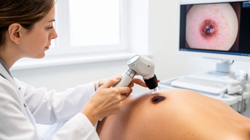
That dual analysis is aimed squarely at the central clinical problem. Melanoma in its early stages can be visually indistinguishable from benign skin moles, and every delay in diagnosis increases the risk that the cancer will metastasize. Current practice places a heavy burden on referral chains: a primary care physician who spots a suspicious lesion typically sends the patient to a dermatologist, who examines the lesion with a dermoscope, a magnifying instrument that reveals subsurface structures, and then performs a biopsy to confirm or rule out cancer. Each step in that chain introduces waiting time, and waiting time is precisely what melanoma patients cannot afford.
The field of AI-assisted dermatology is already moving. In 2024, United States regulators approved an AI-powered tool for evaluating skin lesions and providing information to primary care physicians, a milestone that signaled official recognition of the technology’s potential. Still, building an algorithm that can genuinely match the diagnostic skill of an experienced dermatologist remains an elusive goal. Skin lesions vary enormously in appearance across skin tones, ages, and anatomical locations, and the boundary between benign and malignant features is subtle enough that even expert annotators sometimes disagree. The WPI team is explicit about its intentions: they do not expect AI to replace dermatologists. Instead, they envision AI as a triage instrument embedded in everyday clinical practice.
Garcia Gonzalez, a Fulbright scholar who also serves as a research associate on the electrical engineering faculty at the Technological University of Panama, describes the practical vision behind the project. The team is aiming to create a handheld tool that primary care physicians could use during a routine visit to image a suspicious lesion and receive an immediate AI assessment. Such a device could help doctors decide which patients genuinely need referral to dermatologists, potentially compressing the diagnostic timeline and leading to earlier detection of skin cancer. In regions with limited access to specialists, that kind of decision support could be transformative.
Training an AI system to see what dermatologists see requires enormous volumes of data. In the ISIC challenge, each participating team received a dataset of 5,294 anonymous images to train their models and a second dataset of 479 anonymous images on which to demonstrate performance. The competition’s organizers used the Dice coefficient as the ranking metric, a statistical measure of how closely each model’s diagnoses matched those of human experts. TwinSight earned a score of 0.662 on that measure, placing it third in a field of 181 teams drawn from academia and industry. The score reflects the model’s ability to segment and classify lesion imagery in agreement with expert judgment, a demanding standard given the visual ambiguity of early melanoma.
The WPI team supplemented the competition data with a validation dataset of 187 images obtained from a hospital in Florida, using it to check how the model behaved on imagery outside the challenge’s training distribution. That kind of independent validation is a critical step in machine learning research, because models can sometimes overfit to the statistical quirks of a single dataset and then falter on new data. The team is now collaborating with dermatologists in Florida to develop and test TwinSight further, with the goal of moving the system toward potential use in real clinical settings.
The device the team envisions is deliberately simple in concept: a handheld unit tethered to a laptop or computer tablet. A clinician would place the device on a patient’s skin to collect a finely detailed photograph, which would then be analyzed by the AI in real time. The immediate feedback loop is the point. Rather than waiting days or weeks for a specialist appointment, a physician could receive an instant computational assessment of the lesion’s visual characteristics, informing the referral decision on the spot. Huang frames the ultimate goal in direct terms: helping doctors make faster and more confident decisions when examining patients. Achieving that, he says, requires AI tools to continue evolving, and the team’s third-place finish in a fiercely competitive international field suggests that the evolution is well underway.
Subject of Research: AI-based melanoma detection using dual deep-learning architectures on dermoscopic skin images
Article Title: Worcester Polytechnic Institute developing AI tool to spot skin cancer
Article References: Worcester Polytechnic Institute developing AI tool to spot skin cancer. Original publication (Bibliographic details unavailable.)
Image Credits: AI Generated
DOI: Not provided
Keywords: melanoma, artificial intelligence, skin cancer, deep learning, Worcester Polytechnic Institute, medical imaging, Worcester, Polytechnic, Institute, developing, tool, spot
Cite Scienmag News
Nathaniel Bowman. (October 17, 2026). AI Model From WPI Takes Third Place in Global Race to Spot Melanoma Early. Scienmag. https://scienmag.com/ai-model-from-wpi-takes-third-place-in-global-race-to-spot-melanoma-early/
Nathaniel Bowman. "AI Model From WPI Takes Third Place in Global Race to Spot Melanoma Early." Scienmag, 17 October 2026, https://scienmag.com/ai-model-from-wpi-takes-third-place-in-global-race-to-spot-melanoma-early/. Accessed 17 October 2026.
Nathaniel Bowman. "AI Model From WPI Takes Third Place in Global Race to Spot Melanoma Early." Scienmag. October 17, 2026. https://scienmag.com/ai-model-from-wpi-takes-third-place-in-global-race-to-spot-melanoma-early/


