


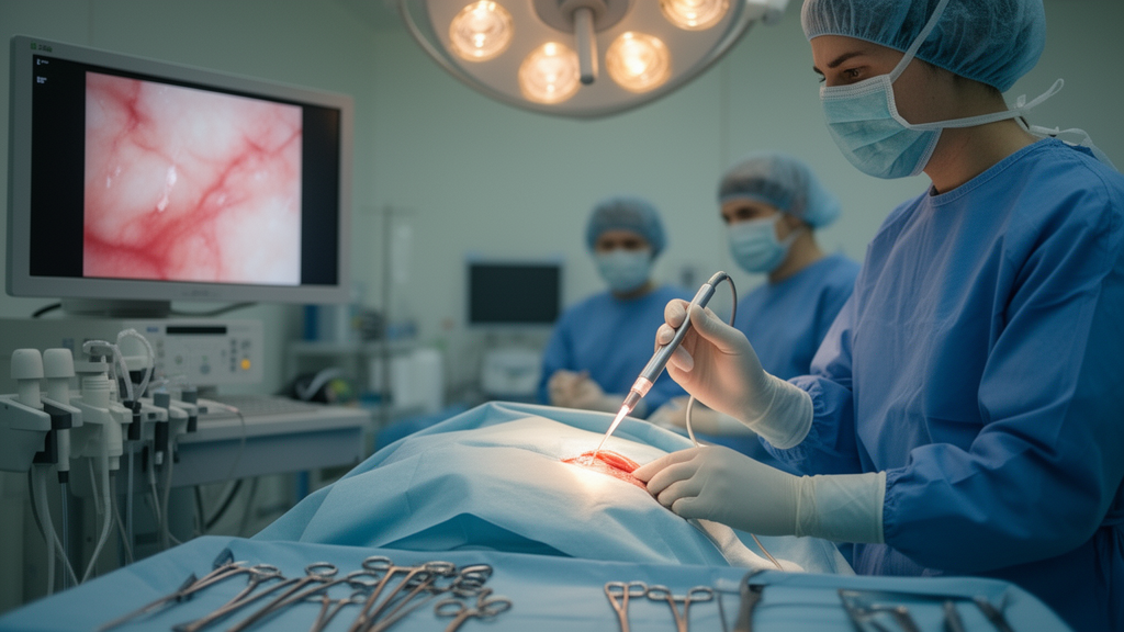
Every year, hundreds of thousands of women undergo breast-conserving surgery to remove a tumour while preserving as much healthy tissue as possible. Yet in roughly one in five of these operations, the surgeon unknowingly leaves cancer cells behind at the edge of the excision, forcing a second procedure weeks later. A comprehensive review now argues that a laser-based imaging technique capable of reading tissue at cellular resolution in real time could dramatically cut those re-operations — and that artificial intelligence and surgical robots are the keys to making it work in the operating theatre.
The review, published in Frontiers in Medical Engineering by researchers at Imperial College London’s Hamlyn Centre for Robotic Surgery and Charing Cross Hospital, examined confocal fluorescence microscopy (CFM), an optical biopsy technique that captures microscopic images of fresh tissue without the need for fixation, sectioning or staining protocols that take hours in a pathology lab. The team screened 1,382 articles published between January 2013 and December 2024 and distilled them into 28 studies that met their criteria, focusing on how well different CFM systems could distinguish malignant from healthy breast tissue against the gold standard of hematoxylin and eosin histopathology.
The technology works by exploiting a fundamental optical principle. A laser illuminates a single point on the tissue surface, and a pinhole aperture ensures that the detector collects light only from that focused spot, rejecting all out-of-focus light from surrounding structures. The result is a razor-thin optical slice through the sample — sharp, cellular images of tissue that has not been physically cut. By scanning the laser spot point-by-point in raster or spiral patterns, the system builds up a two-dimensional image in seconds. Because most breast cancers arise from the epithelial lining of ducts and lobules, fluorescent dyes that bind to cell nuclei make malignant structures stand out with striking contrast.
The review splits CFM systems into two camps. Bench-top systems, such as the commercial VivaScope 2500 and the Histolog Scanner, deliver the highest resolution — down to one micron laterally — and diagnostic accuracy ranging from 83 to 99.6 per cent. In one landmark study, a confocal strip mosaicking microscope imaged a 10-by-10-millimetre area of fresh breast tissue in just one minute with sub-cellular resolution. The Histolog Scanner, which uses a 488-nanometre laser and a proprietary dip stain, can scan all six faces of a lumpectomy specimen in 13 seconds, and retrospective review suggested it could have identified missed tumour margins in up to 75 per cent of cases requiring re-excision. But their size and operational complexity confine them to the pathology bench rather than the live surgical field.
Fibre-based systems take the opposite approach. Flexible fibre bundles act as miniaturised endoscopes that can be inserted into endoscope working channels or surgical cavities, enabling real-time in-situ imaging with accuracy up to 94 per cent. The Cellvizio probe-based confocal laser endomicroscopy system, for example, achieves lateral resolution of one micron over a 240-by-240-micron field of view at 12 frames per second. In a pilot study of 50 patients, topical acriflavine staining allowed pathologists and surgeons to distinguish neoplastic from normal breast tissue within three minutes. A newer line-scan CLE system developed by the Imperial team pushes the frame rate to 120 frames per second — ten times faster than conventional systems — and uses methylene blue, one of the few FDA-approved dyes suitable for in-vivo human application.
The most ambitious frontier involves marrying CFM with surgical robotics. The CRUK-EPSRC-funded MAMMOBOT project has developed a millimetre-scale steerable soft robot that navigates the mammary ducts through the nipple, carrying a flexible endomicroscope in its hollow lumen to perform virtual histology of the ductal system. Feasibility studies showed the line-scan CLE could pass through MAMMOBOT’s channel and identify features corresponding to normal ducts, ductal carcinoma in situ and invasive ductal carcinoma at sub-cellular scale. Robotic scanning systems have also been designed to stabilise the probe against tissue, generate mosaicked imaging fields of up to 14 square millimetres, and reduce the manual variability that plagues hand-held scanning.
Artificial intelligence is the second transformative force. Deep learning models — particularly convolutional and recurrent neural networks — have been trained to segment epithelial structures, stromal boundaries and tumour margins in grayscale confocal images with high precision. A decision tree model validated against expert pathologist-confirmed histopathology achieved 92 per cent accuracy in classifying invasive ductal carcinoma and 96 per cent for ductal carcinoma in situ. Generative adversarial networks are being used to convert grayscale fluorescence images into virtual H&E-like images that resemble the pink-and-purple slides clinicians are accustomed to reading, easing the interpretability gap. AI-driven mosaicking algorithms can stitch thousands of confocal tiles into seamless wide-area maps within minutes, approximating the diagnostic breadth of traditional histology while preserving cellular detail.
Yet the review is candid about the obstacles. The shallow imaging depth of around 100 microns may be insufficient given that surgical guidelines recommend one to two millimetre clear margins. Surgeon interpretation of confocal images was less consistent than pathologist interpretation, with accuracy ranging from 83 to 98 per cent compared to near-perfect 99.6 per cent for pathologists in one study. Many of the fluorescent dyes used for nuclear contrast — acriflavine, proflavine — are mutagenic and not FDA-approved for human use. Bench-top systems can cost more than 200,000 US dollars. And the histological diversity of breast cancer — lobular carcinoma often presents with diffuse infiltration, while ductal carcinoma in situ can be multifocal — means validation across the full spectrum of subtypes remains incomplete, with rarer pathologies collectively accounting for less than two per cent of analysed specimens.
The authors frame the current moment as an inflection point. CFM sits at the intersection of AI-driven diagnostic support, robotic-assisted imaging and virtual histopathology — developments that are no longer distant ambitions but are entering clinical research pipelines. The next steps they identify include developing clinically compatible contrast agents, assembling diverse annotated confocal image datasets for AI training, designing interoperable systems that integrate with existing surgical workflows, and executing multi-institutional trials to validate diagnostic performance across tumour subtypes and clinical environments.
If those hurdles are cleared, the implications extend beyond reducing re-excisions. Real-time digital pathology could shift intra-operative care from a reactive paradigm — waiting days for histopathology results — to a guided one in which surgeons see disease at cellular resolution as they operate. For the one in eight women who will develop breast cancer in their lifetime, that could mean fewer second surgeries, better cosmetic outcomes and a fundamentally new way of visualising and acting on disease in the operating theatre.
Subject of Research: Confocal fluorescence microscopy for real-time breast cancer diagnosis: current advances and future perspectives
Article Title: Confocal fluorescence microscopy for real-time breast cancer diagnosis: current advances and future perspectives
Article References: Vyas, K., Ezzat, A., Holford, N., Ramakrishnan, R., & Leff, D. R. (2025). Confocal fluorescence microscopy for real-time breast cancer diagnosis: current advances and future perspectives. Frontiers in Medical Engineering, 3, Article 1607453. https://doi.org/10.3389/fmede.2025.1607453
Image Credits: AI Generated
DOI: 10.3389/fmede.2025.1607453
Keywords: Confocal, fluorescence, microscopy, real-time, breast, cancer, diagnosis, current, advances, future, perspectives, scientific research
Cite Scienmag News
Nathaniel Bowman. (October 16, 2026). Optical Biopsy Could Give Breast Surgeons Real-Time Cancer Margins. Scienmag. https://scienmag.com/optical-biopsy-could-give-breast-surgeons-real-time-cancer-margins/
Nathaniel Bowman. "Optical Biopsy Could Give Breast Surgeons Real-Time Cancer Margins." Scienmag, 16 October 2026, https://scienmag.com/optical-biopsy-could-give-breast-surgeons-real-time-cancer-margins/. Accessed 16 October 2026.
Nathaniel Bowman. "Optical Biopsy Could Give Breast Surgeons Real-Time Cancer Margins." Scienmag. October 16, 2026. https://scienmag.com/optical-biopsy-could-give-breast-surgeons-real-time-cancer-margins/


