


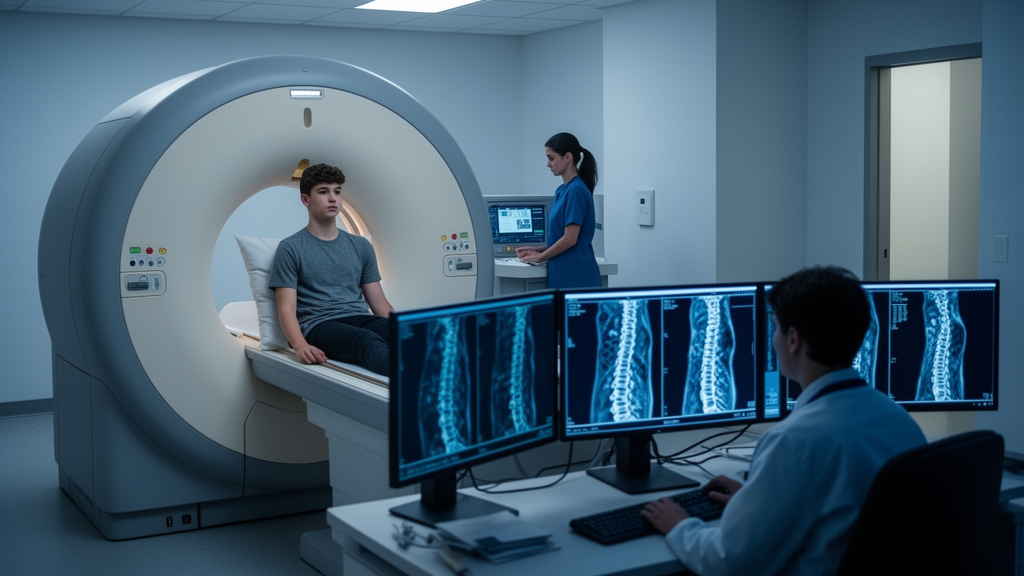
Adolescent athletes who push their spines through years of repetitive loading—gymnasts arching into extreme extension, javelin throwers rotating violently, wrestlers absorbing compressive forces—face a distinctly modern sporting dilemma: the very imaging needed to diagnose their ailing backs relies on ionizing radiation, and repeated exposures across a young lifetime are anything but trivial. A research team at the University of Potsdam has now put one potential radiation-free alternative, upright magnetic resonance imaging, through a rigorous feasibility and reliability test in young athletes with isthmic spondylolisthesis, and the results paint a nuanced picture of promise paired with important caveats.
Isthmic spondylolisthesis is a condition in which a stress fracture of the pars interarticularis—a small bony bridge in the back of each vertebra—allows one vertebral body to slip forward on the one beneath it, most commonly at the L5/S1 junction where the lumbar spine meets the sacrum. The condition affects an estimated four to eight percent of the population by adulthood and shows a strong association with high-level physical activity, particularly sports involving repetitive lumbar extension and rotation under load. In adolescents, the slippage is typically graded on standing X-rays using the Meyerding classification, which quantifies the degree of forward translation from grade I, representing up to twenty-five percent slippage, through grade V, in which the vertebra has slid entirely off the one below.
The clinical gold standard for diagnosing and grading this slippage has long been upright, lateral radiography performed in two planes. But conventional X-rays, while excellent at depicting bone, offer limited information about the soft-tissue neighborhood of the slipped segment: the intervertebral disc, the facet joints, the nerve-root pathways, and the paravertebral muscles. Magnetic resonance imaging can visualize all of these structures in exquisite detail, yet standard MRI scanners image patients lying flat, unloaded—a position in which the spine behaves differently than it does under the compressive forces of standing. Upright MRI, which images patients in a weight-bearing posture, promises to combine the best of both worlds: radiation-free soft-tissue imaging of a loaded, functional spine. Whether that promise survives contact with the measurement precision demanded by clinical decision-making is precisely what the Potsdam team set out to determine.
The researchers retrospectively reviewed imaging data from thirty-eight adolescent athletes, aged roughly eleven to seventeen, in whom isthmic spondylolisthesis had been diagnosed during preparticipation examinations for competitive sports between 2009 and 2019. The athletic backgrounds of the cohort reflected the demands of their disciplines: nine wrestlers, seven canoeists, seven track-and-field athletes, six judoka, and smaller numbers of swimmers, weightlifters, rowers, riders, soccer players, cyclists, and a gymnast. Each participant had undergone lateral standing radiographs and sagittal T2-weighted upright MRI on a low-field 0.25-Tesla scanner, with the MRI performed at an eighty-degree inclination in a standardized weight-bearing position using straps, a knee roll, and arm support to stabilize the body.
From the original cohort, only sixteen athletes—ten female and six male, with a mean age of 13.4 years—produced image pairs that met the study’s strict inclusion criteria: both examinations free of motion artifacts, performed within one year of each other, and clearly depicting the entire lumbar spine and lumbopelvic junction. Nine participants were excluded because the interval between their two examinations exceeded one year, and eight because image quality was insufficient, largely due to motion artifacts accumulated during the roughly ten-minute standing acquisition. All sixteen had slippage at the L5/S1 level, twelve at Meyerding grade I and four at grade II.
On the evaluable images, a single experienced examiner measured five key spinopelvic and lumbar parameters twice, at least seven days apart and blinded to prior results: anterior translation of the slipped vertebra, the lumbosacral joint angle, the lumbar lordosis angle, the sacral slope, and the intervertebral disc height. The team then quantified reliability using a battery of statistical tools. Intraclass correlation coefficients captured relative reliability, while standard error of measurement, test-retest variability, minimal detectable change, and Bland-Altman analysis captured the absolute magnitude of measurement error and any systematic bias between techniques.
The findings revealed a clear hierarchy of precision. Radiographs delivered excellent intra-rater repeatability, with intraclass correlation coefficients ranging from 0.89 for disc height to 0.95 for anterior translation, and test-retest variability between five and fourteen percent. The minimal detectable change for anterior translation on radiographs was a remarkably tight 1.4 millimeters, meaning that only changes exceeding this threshold can be confidently distinguished from measurement noise. Upright MRI, by contrast, showed moderate-to-excellent repeatability for several parameters—sacral slope reached an impressive 0.94, and anterior translation a solid 0.87—but the lumbosacral joint angle proved far less stable, with test-retest variability reaching forty-nine percent and a standard error of measurement of twenty-nine percent. Disc height on upright MRI yielded the weakest correlation coefficient at 0.61. Notably, Bland-Altman analysis detected no systematic measurement error within either modality, suggesting the discrepancies were random rather than directional.
The cross-modality comparison between radiographs and upright MRI proved the study’s most sobering result. Agreement between the two techniques was parameter-dependent and highly variable, with correlation coefficients ranging from 0.53 for disc height to 0.88 for lumbar lordosis. Anterior translation—the parameter clinicians care most about for grading slippage—showed only moderate agreement at 0.61, with a minimal detectable change of 3.2 millimeters between modalities, more than double the radiographic figure. The lumbar lordosis angle, despite its relatively high correlation coefficient, exhibited a systematic bias of minus 4.3 degrees on upright MRI compared to radiographs, a discrepancy the authors attribute in part to differences in positioning: the MRI was acquired at eighty degrees with external stabilization, while radiographs were taken unsupported at a full ninety-degree stance. These findings, the researchers conclude, do not support interchangeability between the two modalities.
The study’s limitations are candidly acknowledged and materially important. The low field strength of the 0.25-Tesla scanner limited spatial resolution compared with the 1.5-Tesla systems the authors recommend for clinical practice, and the roughly ten-minute standing acquisition made young participants vulnerable to motion artifacts—thirteen of thirty-eight upright MRI scans were deemed illegible for this reason. The retrospective design meant that radiographs and MRI scans were separated by an average of 5.2 months, during which adolescent spinal morphology may have changed, and operator-related variability across five radiography and two MRI technicians could not be excluded. Information on radiographic magnification correction was also unavailable, leaving potential uncertainty in linear measurements. With only sixteen participants in the paired analysis, the precision and generalizability of the reliability estimates are inherently constrained, and restricting analysis to evaluable images may have overestimated performance relative to routine clinical imaging.
Nevertheless, the study establishes a meaningful foundation for what may become a significant shift in how young athletes’ spines are monitored. Radiography should remain the reference standard for diagnosing and grading isthmic spondylolisthesis for now, the authors conclude, but upright MRI may serve as a complementary, radiation-free tool for selected patients and selected parameters—particularly where clinicians want to assess the loaded spine while simultaneously visualizing bone marrow edema, disc pathology, facet joint degeneration, stenosis, or paravertebral muscle changes that X-rays simply cannot show. Future prospective studies, the team argues, should employ same-day imaging, standardized and equivalent positioning across modalities, randomized acquisition order, and higher-field upright systems, with examination times kept below ten minutes to accommodate the realities of imaging restless adolescent bodies. Until then, the message for clinicians managing young athletes with back pain is one of cautious optimism: the radiation-free future of spinal imaging is drawing closer, but the numbers say it has not yet arrived.
Subject of Research: Reliability of upright MRI versus radiography for measuring spinopelvic and lumbar parameters in adolescent athletes with isthmic spondylolisthesis
Article Title: Exploratory evaluation of spinopelvic and lumbar parameters in adolescent athletes with isthmic spondylolisthesis: feasibility and reliability of upright MRI compared to radiography
Article References: Cassel, M., Joost, T., Slater, C., Mayer, F., & Engel, T. (2026). Exploratory evaluation of spinopelvic and lumbar parameters in adolescent athletes with isthmic spondylolisthesis: feasibility and reliability of upright MRI compared to radiography. Frontiers in Radiology, 6, Article 1929174. https://doi.org/10.3389/fradi.2026.1929174
Image Credits: AI Generated
DOI: 10.3389/fradi.2026.1929174
Keywords: isthmic spondylolisthesis, upright MRI, adolescent athletes, radiography, spinopelvic parameters, lumbar spine, measurement reliability, Meyerding classification, low-field MRI, radiation-free imaging, spondylolysis, L5/S1
Cite Scienmag News
Ophelia Keating. (October 16, 2026). Upright MRI Shows Promise but Cannot Yet Replace X-Rays for Young Athletes’ Spine Slips. Scienmag. https://scienmag.com/upright-mri-shows-promise-but-cannot-yet-replace-x-rays-for-young-athletes-spine-slips/
Ophelia Keating. "Upright MRI Shows Promise but Cannot Yet Replace X-Rays for Young Athletes’ Spine Slips." Scienmag, 16 October 2026, https://scienmag.com/upright-mri-shows-promise-but-cannot-yet-replace-x-rays-for-young-athletes-spine-slips/. Accessed 16 October 2026.
Ophelia Keating. "Upright MRI Shows Promise but Cannot Yet Replace X-Rays for Young Athletes’ Spine Slips." Scienmag. October 16, 2026. https://scienmag.com/upright-mri-shows-promise-but-cannot-yet-replace-x-rays-for-young-athletes-spine-slips/


