


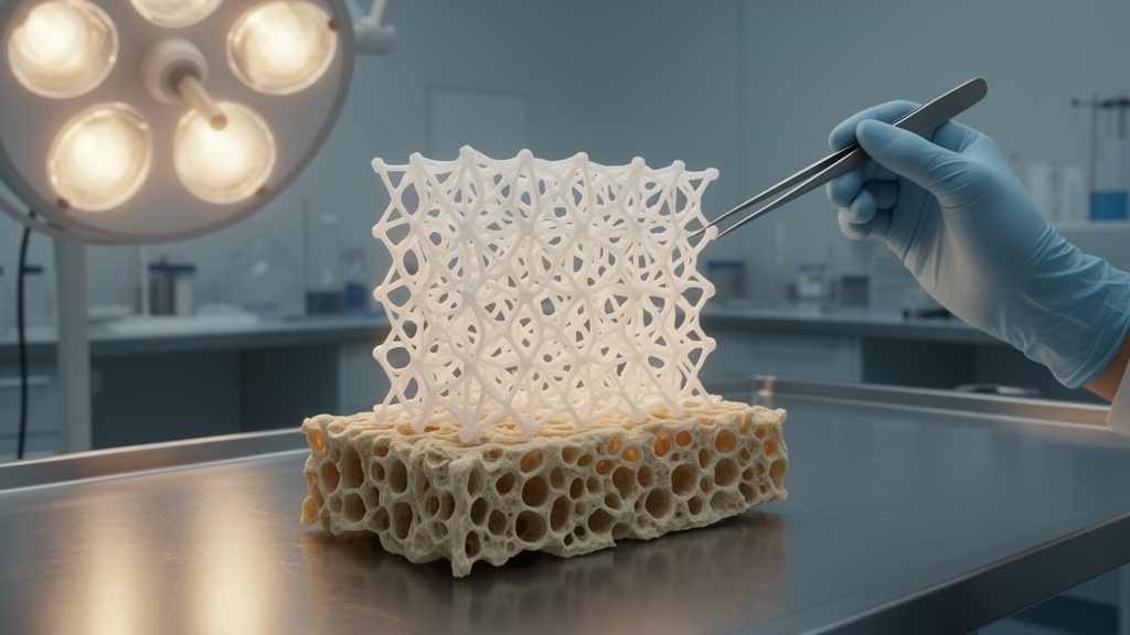
Osteoporosis affects more than 200 million people worldwide and triggers an estimated 8.9 million fragility fractures every year — one fracture roughly every three seconds. For decades, surgeons treating these broken bones have relied on implants that act essentially as passive hardware: titanium plates, screws, and cages that hold fragments in place while the body attempts its own repair. The problem is that in osteoporotic patients, the body’s repair machinery is itself broken. Bone-forming cells lose their vigor, bone-resorbing cells run unchecked, oxidative stress and chronic inflammation poison the local environment, and blood vessel growth falters. The result is delayed healing, failed unions, and implants that loosen long before they should.
A sweeping new mini-review published in Frontiers in Biomaterials Science by researchers from the University of Santiago de Compostela, the Spanish National Research Council, and the Complutense University of Madrid argues that the next generation of implants must do far more than fill a defect. Writing from a translational perspective, Miguel Castro-Rodríguez, Luis Diaz-Gomez, Yolanda Sanz, Carmen Alvarez-Lorenzo, and María Vallet-Regí synthesized evidence on three-dimensional and four-dimensional printed scaffolds specifically engineered for osteoporotic bone, drawing on in vivo preclinical studies and the scarce but growing body of human-oriented work. Their conclusion is striking: passive structural support is often insufficient in osteoporotic bone, and the most promising scaffolds are active therapeutic devices — drug-loaded, immunomodulatory, antioxidant, osteoanabolic, and even stimuli-responsive platforms that treat the disease while rebuilding the tissue.
Three-dimensional printing has become the backbone of this strategy because it allows engineers to control porosity, stiffness, and surface chemistry with remarkable precision, producing patient-specific implants whose internal architecture can be tuned to match the mechanical properties of fragile bone. The review catalogues an impressive range of materials that have shown enhanced osseointegration and bone regeneration in osteoporotic animal models: porous titanium and titanium alloys, tantalum, biodegradable magnesium alloys, calcium phosphate ceramics, bioactive glass, polycaprolactone, polylactic acid, polyetheretherketone, and hybrid hydrogel-integrated composites. But the real excitement lies in what these scaffolds carry. Functional platforms have been loaded locally with simvastatin, zoledronic acid, alendronate, teriparatide, bone morphogenetic proteins, platelet-rich plasma, strontium, magnesium, lanthanum, cerium, icaritin, and a growing arsenal of antioxidant and immunomodulatory agents.
Some of the most technically sophisticated designs illustrate how architecture and biology can be fused. A marine sponge-inspired titanium alloy scaffold, dubbed a cell climbing frame, uses nested macro- and micropores to lower elastic modulus, improve blood penetration, and recruit and rejuvenate the patient’s own bone marrow stromal cells through embedded E7 peptides and exosomes — accelerating bone repair and delaying surrounding bone loss in osteoporotic rats. A 3D-printed tantalum scaffold enhanced repair of osteoporotic defects by modulating the SDF-1α/CXCR4 signaling axis, suggesting that scaffold geometry itself can influence endogenous cell recruitment and molecular repair pathways. Meanwhile, a fishnet-inspired scaffold combining mesh-like electrospun membranes with strontium-substituted hydroxyapatite-enriched polycaprolactone and silk fibroin nanofibers achieved near-complete repair of osteoporotic cranial defects within just eight weeks, attributed to combined angiogenic, osteogenic, and anti-osteoclastic effects.
Local drug delivery has emerged as a dominant strategy because it concentrates osteoanabolic or antiresorptive drugs at the defect while reducing systemic exposure. Photocurable mesoporous bioactive glass scaffolds coordinated with teriparatide — the 1-34 fragment of parathyroid hormone — bidirectionally regulated bone homeostasis by promoting formation and inhibiting resorption. Dual-controlled release systems based on PLGA microspheres embedded in chitosan and nano-hydroxyapatite hydrogel inks produced sustained biphasic teriparatide delivery and significantly improved bone volume fraction and trabecular thickness in ovariectomized rat calvarial defects. Sequential release platforms are particularly attractive because bone healing requires coordination of immune modulation, osteoclast inhibition, angiogenesis, and osteogenesis across distinct phases. One β-tricalcium phosphate scaffold sequentially releasing interleukin-4 and alendronate first promoted M2 macrophage polarization and subsequently inhibited osteoclast activity while supporting osteogenic differentiation, resulting in superior regeneration in an osteoporotic rat femoral defect model.
The emerging frontier is four-dimensional printing — scaffolds whose properties evolve over time in response to internal or external cues. These systems exploit pathological features of the osteoporotic microenvironment, including acidity, oxidative stress, and chronic inflammation, to achieve controlled therapeutic delivery. Strontium carbonate-polydopamine scaffolds released strontium ions under acidic conditions, suppressing osteoclast activity and promoting M2 macrophage polarization. Melatonin-loaded zeolitic imidazolate framework nanoparticles in gelatin methacryloyl hydrogel coatings provided pH-responsive release of zinc ions and melatonin, reducing oxidative stress and restoring mitochondrial function. Temperature-responsive collagen coatings containing free 4-octyl itaconate and BMP-9-loaded particles remained fluid during scaffold coating and gelled at physiological temperature after implantation, delivering rapid anti-inflammatory release followed by sustained osteogenic stimulation. External stimuli add another dimension: near-infrared-responsive MXene-polylactic acid-icaritin scaffolds employed mild photothermal activation to trigger drug release and accelerate bone repair in a rat vertebral fusion model, while a tannic-acid-based photothermal hydrogel coating on 3D-printed titanium scaffolds incorporated Prussian blue, quercetin, osteogenic growth peptide, and SDF-1α to promote cell recruitment, ROS scavenging, anti-senescence effects, and M2 macrophage polarization in vivo.
Despite this impressive preclinical progress, the review delivers a sobering reality check: true human clinical evidence remains scarce. Most human-oriented work is limited to finite element modeling, patient-specific implant design, biomechanical testing, cadaveric studies, or imaging phantoms rather than therapeutic trials. Anatomical contour titanium oblique lumbar interbody fusion cages with porous lattice structures and embedded fixation screws have been optimized using finite element analysis and weight topology optimization based on osteoporotic endplate morphology, showing improved stability and lower endplate stress. A hybrid titanium 3D-printed suture anchor with an expandable wing mechanism demonstrated higher failure strength than a commercially available solid anchor in osteoporotic bone models, with in vivo implantation in pigs showing bone ingrowth along the anchor contour comparable to the commercial device. Yet these are design and validation studies, not clinical effectiveness trials.
Several ongoing and recent clinical studies nonetheless offer a glimpse of what translation might look like. A prospective observational cohort study is evaluating 3D-printed porous titanium alloy cages compared with conventional PEEK cages in osteoporotic patients undergoing posterior lumbar interbody fusion, with primary outcomes including screw loosening and fusion rates at six months. A related multicenter prospective cohort study investigates biomechanically optimized 3D-printed titanium cages specifically designed to match the elastic modulus of osteoporotic bone, aiming to reduce cage subsidence and improve fusion outcomes. Beyond spinal indications, a randomized controlled trial is assessing patient-specific, resorbable polycaprolactone scaffolds for maxillary and mandibular bone regeneration, comparing their osteogenic potential with conventional guided bone regeneration techniques using PTFE membranes and xenografts. These studies underscore growing clinical interest in improving osseointegration, achieving better biomechanical compatibility, and enabling patient-specific treatments — but most current evidence derives from observational designs or early-phase trials with relatively small cohorts.
The review identifies several formidable challenges that must be overcome before widespread clinical adoption. Osteoporotic bone repair is multifactorial, so a scaffold that only stimulates osteogenesis may fail if inflammation, oxidative stress, osteoclast activity, or poor mechanical fixation are not simultaneously addressed. Model standardization is another hurdle: most in vivo studies rely on ovariectomized rodent models with calvarial or femoral defects, which do not fully reproduce human fragility fractures, spinal fusion, joint arthroplasty, or elderly multimorbidity. Large-animal validation, mechanically loaded models, aged animals, and direct comparisons with current clinical standards are needed. Clinical manufacturability poses its own difficulties, requiring consistent printing quality, sterilization, quality control, and regulatory documentation — and for four-dimensional systems, the challenge is greater still because the device may combine scaffold, drug, biologic, and stimulus-responsive behavior in a single construct.
The trajectory, however, is unmistakable. Three-dimensional-printed scaffolds have advanced from inert porous supports to bioactive, drug-delivering, immunomodulatory, and antioxidant platforms, and four-dimensional stimuli-responsive systems represent the next stage, offering temporal and microenvironment-adaptive control over repair. The field remains predominantly preclinical, but the convergence of advanced manufacturing, precision drug delivery, and deepening understanding of the osteoporotic microenvironment is setting the stage for a new era in which the implant itself becomes a treatment. Translation will depend on rigorous long-term in vivo validation, large-animal studies, standardized outcomes, scalable manufacturing, reproducible fabrication, long-term safety data, and early-phase clinical trials in osteoporotic patients. If those hurdles can be cleared, the humble bone scaffold may soon evolve from passive hardware into an active, adaptive therapy — one that treats osteoporosis while it rebuilds the bone it holds together.
Subject of Research: 3D- and 4D-printed scaffolds for osteoporotic bone regeneration: translational insights from in vivo preclinical and human-oriented studies
Article Title: 3D- and 4D-printed scaffolds for osteoporotic bone regeneration: translational insights from in vivo preclinical and human-oriented studies
Article References: Castro-Rodríguez, M., Diaz-Gomez, L., Sanz, Y., Alvarez-Lorenzo, C., & Vallet-Regí, M. (2026). 3D- and 4D-printed scaffolds for osteoporotic bone regeneration: translational insights from in vivo preclinical and human-oriented studies. Frontiers in Biomaterials Science, 5, Article 1937864. https://doi.org/10.3389/fbiom.2026.1937864
Image Credits: AI Generated
DOI: 10.3389/fbiom.2026.1937864
Keywords: D-printed, scaffolds, osteoporotic, bone, regeneration, translational, insights, vivo, preclinical, human-oriented, studies, scientific research
Cite Scienmag News
Denise Maddox. (October 16, 2026). Printed Scaffolds That Heal Osteoporotic Bone Are Moving From Lab to Clinic. Scienmag. https://scienmag.com/printed-scaffolds-that-heal-osteoporotic-bone-are-moving-from-lab-to-clinic/
Denise Maddox. "Printed Scaffolds That Heal Osteoporotic Bone Are Moving From Lab to Clinic." Scienmag, 16 October 2026, https://scienmag.com/printed-scaffolds-that-heal-osteoporotic-bone-are-moving-from-lab-to-clinic/. Accessed 16 October 2026.
Denise Maddox. "Printed Scaffolds That Heal Osteoporotic Bone Are Moving From Lab to Clinic." Scienmag. October 16, 2026. https://scienmag.com/printed-scaffolds-that-heal-osteoporotic-bone-are-moving-from-lab-to-clinic/


