


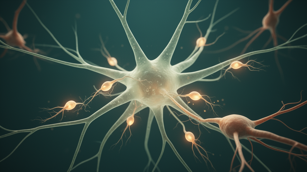
Deep in the dentate gyrus of the hippocampus, a remarkable process continues throughout life: new neurons are born from neural stem and progenitor cells (NSPCs). But these stem cells do not act alone. They are embedded in a niche of supporting cells, chief among them astrocytes, the star-shaped glial cells long known for their housekeeping duties. A new study published in Frontiers in Cellular Neuroscience now shows that astrocytes are not static supporters but active, stage-specific instructors of neurogenesis, and that their instructional power changes dramatically as they themselves mature during early postnatal development.
Astrocytes are essential regulators of brain development. They coordinate synapse formation, recycle neurotransmitters, provide metabolic support, and influence the behavior of radial glia-like neural stem cells. In the developing cerebral cortex, astrogenesis follows the primary wave of neurogenesis, with embryonic radial-glia-like NSCs transitioning into proliferating progenitors that fill both the neuronal and glial compartments. Newly born astrocytes then undergo a gradual postnatal transition, progressively elaborating their complex morphology in direct response to developing neuronal circuits, shifting their gene expression programs, and acquiring mature physiological functions. In most brain regions these developmental processes are completed, persisting only in the adult neurogenic niches: the dentate gyrus and the subependymal zone of the lateral ventricle.
The dentate gyrus is formed from late embryonic stages onward and proceeds into the first postnatal weeks. Neurogenesis starts first, astrogenesis begins after birth, and the two processes continue to run in parallel lifelong. At postnatal day 3 (p3), the dentate gyrus is dominated by extensive proliferation of radial glia-like NSCs that have generated very little neuronal and astrocytic progeny so far. By p7, the NSC pool has decreased while neuronally determined NSPCs expand and the first astrocytes appear in the superficial layers. Between p10 and p14, NSCs continue to decline as the neuronal compartment expands steadily and astrocytes progressively occupy the deeper layers. Notably, developing astrocytes follow the same outside-in pattern described for granule neurons, meaning they colonize the dentate gyrus only after neurons are already present. As a consequence, developing astrocytes are born into an environment shaped by existing neurons, and their morphological expansion occurs in parallel with extensive synaptogenesis and circuit formation.
Astrocytes influence neurogenesis through a wide range of contact-mediated and secreted signals. Classical astrocyte-derived molecules, including thrombospondins, HEVIN, SPARC, glypicans, and cholesterol-associated lipoproteins, promote synapse formation and maturation. Astrocytes also release cytokines and growth factors that modulate NSC proliferation and differentiation. In addition to these protein-based cues, metabolites such as lactate, ketone bodies, and lipids have emerged as important mediators of neuron-glia communication, influencing NSC fate decisions, neuronal differentiation, and synapse formation. However, the contribution of metabolites to neurogenesis is not fully understood. Comparisons across CNS regions have shown that astrocytes from non-neurogenic regions, such as the adult spinal cord, lack many pro-neurogenic cues, indicating that astrocyte identity and secretome are tightly linked to developmental stage and local environment.
To define how hippocampal astrocytes mature molecularly across postnatal development, and how their developmental stage shapes the signals they release, the researchers established a method to isolate astrocytes from key developmental time points: p3, p10, and p14, corresponding to progenitor-like, early-mature, and mature astrocytes in vivo. Using magnetic activated cell sorting with ACSA-2 microbeads, they purified hippocampal astrocytes from C57BL/6J mice at these ages and cultured them for seven days in serum-free medium. Serum exposure can substantially alter astrocyte morphology and transcriptional states, so serum-free conditions were used to preserve the characteristic ramified morphology and prevent serum-induced reactive changes, maintaining an in vivo-like state. Astrocyte conditioned medium (ACM) was collected throughout the culture period by exchanging half of the medium every other day.
The functional assays were striking. Adult hippocampal NSPCs isolated from the dentate gyrus of adult mice were forced to differentiate by withdrawal of growth factors, and instead of standard differentiation medium they were cultured in ACM from p3, p10, or p14 astrocytes for five days. After fixation, immunofluorescence staining against markers for mature neurons (MAP2), immature neurons (DCX), and astrocytes (GFAP) revealed a dramatic stage-dependent shift. ACM from p3 astrocytes resembled the negative control condition, giving rise to mostly GFAP-positive cells (74.20 percent) and few neurons (9.08 percent). In contrast, ACM from p10 and p14 astrocytes substantially increased neuronal differentiation and maturation. P10 ACM yielded 51.79 percent MAP2-positive neurons and 35.92 percent GFAP-positive astrocytes, while ACM from p14 astrocytes produced the highest MAP2-positive fraction (62.85 percent) and even fewer GFAP-positive astrocytes (23.72 percent). Notably, the proportion of DCX-positive immature neurons remained unchanged across all conditions, suggesting that the astrocyte-derived factors act on later steps of neuronal differentiation and maturation rather than on the initial commitment to the neuronal lineage.
To characterize the transcriptional programs underlying postnatal astrocyte identity, the team performed bulk RNA sequencing on directly isolated hippocampal astrocytes at p3, p10, and p14, processing freshly isolated cell pellets immediately to capture their stage-specific transcriptional state without culture-induced alterations. Astrocytes shared a large core transcriptome consisting of 11,159 genes across all three stages, and Gene Ontology enrichment analysis revealed persistent biosynthetic and astrocytic-lineage programs, including RNA metabolism, protein localization, mitochondrial organization, phospholipid metabolism, alongside neural-related categories such as synapse organization and regulation of neurogenesis and astrogenesis. Core astrocyte markers, including Atp1b2, Gfap, Aqp4, Aldh1l1, Aldoc, Apoe, Slc1a2, and Slc1a3, were highly expressed across all samples, confirming robust astrocyte enrichment. Stem cell-associated genes such as Vimentin, Sox2, Pax6, and Prom1 were consistently low, although Nestin was elevated at p3, reflecting the expected presence of stem cells and glial progenitor cells at this early stage.
Stage-enriched differential expression analysis revealed clear developmental distinctions. While p3 astrocytes were defined by early-response and cell cycle-associated genes, including Junb, Fos, Egr2, Zpf36, Ccna1, and Fscn1, p14 astrocytes exhibited a more mature transcriptional profile, expressing genes such as Ogdhl, Maob, Etnppl, Ezr, Gas6, and Slc24a4. Interestingly, p10 astrocytes showed comparatively few stage-enriched differentially expressed genes and appear to represent an intermediate transcriptional state. Consistent with their proliferative identity, p3 astrocytes were significantly enriched for mitotic and DNA replication pathways. In contrast, p10 and p14 astrocytes shared many biological processes, including lipid metabolic processes, synapse-related pathways, and oxidative metabolism. However, p10 astrocytes showed greater enrichment for lipid and fatty acid metabolic programs and synaptogenic pathways, whereas p14 astrocytes exhibited higher enrichment for oxidative phosphorylation, aerobic respiration, and small molecule catabolism. Together, these data reveal a coordinated transcriptional progression in which astrocytes transition from a proliferative state at p3, through a metabolically shifting and synaptogenic phase at p10, to a mature mitochondrial and neuro-protective state by p14.
The researchers also compared directly isolated astrocytes with their cultured counterparts. Principal component analysis revealed a clear separation between the direct and the cultured astrocytes at all stages. While the directly isolated astrocytes separated well according to the time point of isolation, the cultured astrocytes appeared rather intermingled, indicating that the in vitro environment induces a transcriptional state that diverges from in vivo astrocyte maturation. Directly isolated astrocytes upregulated physiological and metabolic transporter genes such as Slc7a10, Slco1c1, and Grm3, and showed strong enrichment for regulation of synapse structure or activity, synapse assembly, and axon guidance. In contrast, cultured astrocytes were characterized by a prominent upregulation of core stress-adaptation, antioxidant, and cell-cycle checkpoint genes, including Hmox1, Cdkn1a, Srxn1, and Txnrd1, with their broader transcriptional signature dominated by pathways related to chromosome segregation, DNA replication, and nuclear division. Nevertheless, cultured astrocytes continued to express stage-defining genes characteristic of their in vivo counterparts, suggesting that intrinsic transcriptional and epigenetic memory persists for at least a week in culture. Without the appropriate environmental cues, however, this memory was insufficient to maintain normal maturation, leading to the upregulation of proliferative and stress-adaptive pathways.
Perhaps the most exciting findings came from the metabolomic profiling. Since transcriptomics provide only indirect proof of what astrocytes actually release, the team directly compared the secreted metabolites by performing untargeted metabolomic profiling of ACM collected from p3 and p14 astrocytes across four mass-spectrometry modes using high-resolution mass spectrometry coupled to liquid chromatography. Of the 354 detected metabolites, 101 showed at least a two-fold enrichment relative to control medium, ensuring that only metabolites actively secreted by the astrocytes were retained for analysis. From those 101 metabolites, 91 were enriched exclusively in p3 ACM and 10 were enriched exclusively in p14 ACM. Focusing on these 10 factors, the researchers selected arachidonic acid (AA), eicosapentaenoic acid (EPA), L-DOPA, and cholesterol sulfate (CS) for functional testing in adult NSPC cultures. The results were remarkable. The addition of EPA, L-DOPA, and CS at their highest concentrations induced neuronal differentiation at levels comparable to standard NSPC differentiation medium, indicating that all three metabolites alone are potent enough to drive neuronal differentiation from NSPCs. The addition of 10 micromolar AA even exceeded the NSPC’s neurogenic capacity, resulting in 56.4 percent MAP2-positive neurons while reducing GFAP-positive cells to 22.5 percent. The combination of EPA (5 micromolar), AA (10 micromolar), and L-DOPA (50 micromolar) was the strongest inducer of neuronal differentiation, producing 65.7 percent MAP2-positive neurons and only 16.1 percent GFAP-positive astrocytes, outperforming the premixed NSPC differentiation medium by more than 15 percent.
These observations support a model in which early astrocytes provide broad biosynthetic support, whereas more mature astrocytes provide specialized secreted cues capable of modulating hippocampal differentiation. The enrichment of polyunsaturated fatty acids in p14 ACM is consistent with evidence showing that omega-3 and omega-6 PUFAs modulate NSPC behavior and neuronal differentiation. In particular, omega-3s such as docosahexaenoic acid and EPA promote neuronal differentiation by downregulating Hes1 and upregulating activator-type bHLH transcription factors including Hes6, NeuroD, and Mash1, thereby enhancing MAP2 and cell cycle inhibitors like p21 and p27. Beyond these omega-3 mechanisms, both DHA and the omega-6 PUFA AA increase the number of neurogenic NSCs during postnatal development. While classical work suggests that AA primarily supports NSC maintenance, the present data indicate that AA promotes neuronal differentiation and maturation, pointing to a context-dependent role of AA in hippocampal development. In addition, cholesterol sulfate, also enriched at p14, exerts neuroprotective and anti-apoptotic effects, potentially via metabolic support, while catecholamine-related metabolites like L-DOPA modulate oxidative stress responses and dopaminergic lineage specification.
In addition to metabolic cues, classical morphogen pathways may contribute to the proneurogenic effects of the maturing astrocyte secretome. Astrocyte-derived Wnt/beta-catenin signaling is a well-established regulator of adult hippocampal neurogenesis, acting primarily on early steps of neurogenesis, including neuroblast proliferation and neuronal fate commitment. In the transcriptomic dataset, Wnt3 and Wnt3a were expressed at or below the detection threshold in both directly isolated and cultured astrocytes, suggesting that these ligands are unlikely to mediate the pro-maturational effects observed here. Notably, however, Wnt7a and Wnt7b were abundantly expressed in the astrocyte preparations across all developmental stages. Interestingly, expression of Wnt7a peaks in p10 astrocytes, the synaptogenic window during which astrocyte maturation and circuit formation proceed in parallel. Previous work identified an important role for Wnt7a in neuronal differentiation and maturation via beta-catenin-neurogenin 2 signaling and regulation of dendritic development of newborn granule neurons in the dentate gyrus. Furthermore, Wnt7-family signaling regulates axonal remodeling and synapse formation, making Wnt7a a likely contributor to neuronal maturation.
The authors acknowledge several limitations. First, the astrocytes used for bulk RNA sequencing contain a mixture of astrocytes in different developmental stages. While at p3 they harvest mostly NSPCs, p10 and p14 contain both more mature superficial astrocytes and less mature deep layer astrocytes. Second, because six to eight animals were pooled per age group to obtain sufficient cell numbers, the analyses capture population-level averages and cannot resolve heterogeneity between individual animals or between astrocyte subpopulations. Third, the ACM used for the functional assays was collected from astrocytes maintained in culture for seven days, and although cultured astrocytes retain stage-specific transcriptional signatures, culture induces proliferative and stress-adaptive programs that differ from the in vivo environment. Consequently, the data demonstrate that stage-dependent signals promoting neuronal maturation are present in the hippocampal astrocyte secretome, although the approach cannot determine which specific astrocyte subpopulations produce them. Future single-cell and spatial transcriptomic approaches would be required to resolve which astrocyte subsets produce the pro-neurogenic cues identified here.
Overall, the findings position astrocytes as active instructors of postnatal brain development. Rather than serving solely as supportive elements, astrocytes shape both their own maturation program and their direct environment through a combination of transcriptional programs, metabolic activity, and secreted cues. By supplying factors that influence NSC behavior and neuronal differentiation, astrocytes help guide the trajectory of hippocampal circuit formation. These insights reveal a developmental framework in which astrocytes progressively refine their regulatory repertoire, suggesting that shifts in glial states and metabolism represent key mechanisms guiding neuronal maturation and refinement. The identification of a combination of metabolites that accumulates as astrocytes mature and acts as potent drivers of neuronal differentiation opens new avenues for understanding how the metabolic microenvironment of the neurogenic niche is established, and potentially for harnessing these cues in regenerative approaches aimed at promoting neurogenesis in the adult brain.
Subject of Research: Astrocyte maturation creates a stage-specific niche that instructs hippocampal neurogenesis
Article Title: Astrocyte maturation creates a stage-specific niche that instructs hippocampal neurogenesis
Article References: Astrocyte maturation creates a stage-specific niche that instructs hippocampal neurogenesis. (n.d.). https://doi.org/10.3389/fncel.2026.1966983
Image Credits: AI Generated
DOI: 10.3389/fncel.2026.1966983
Keywords: Astrocyte, maturation, creates, stage-specific, niche, instructs, hippocampal, neurogenesis, scientific research
Cite Scienmag News
Cassandra Pierce. (October 16, 2026). Astrocyte Maturation Creates a Stage-Specific Niche That Instructs Hippocampal Neurogenesis. Scienmag. https://scienmag.com/astrocyte-maturation-creates-a-stage-specific-niche-that-instructs-hippocampal-neurogenesis/
Cassandra Pierce. "Astrocyte Maturation Creates a Stage-Specific Niche That Instructs Hippocampal Neurogenesis." Scienmag, 16 October 2026, https://scienmag.com/astrocyte-maturation-creates-a-stage-specific-niche-that-instructs-hippocampal-neurogenesis/. Accessed 16 October 2026.
Cassandra Pierce. "Astrocyte Maturation Creates a Stage-Specific Niche That Instructs Hippocampal Neurogenesis." Scienmag. October 16, 2026. https://scienmag.com/astrocyte-maturation-creates-a-stage-specific-niche-that-instructs-hippocampal-neurogenesis/


