


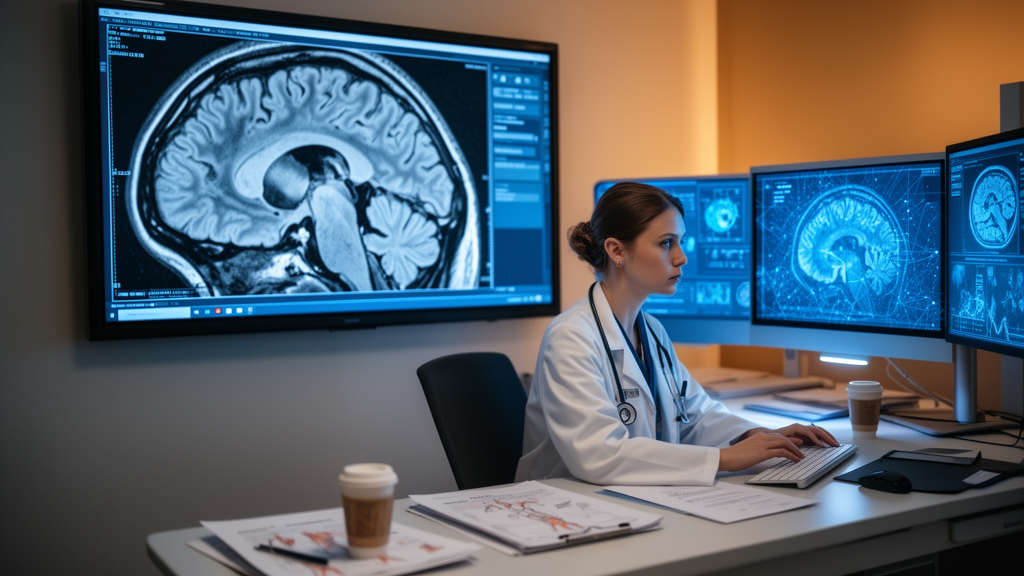
Machine learning models can now outline a brain tumor on an MRI scan with an accuracy that would have seemed like science fiction a decade ago. A sweeping new systematic review, published in Frontiers in Computational Neuroscience, has audited the entire field — 213 studies published between January 2018 and March 2026 — and delivered a verdict that is as sobering as it is statistically precise: the benchmark scores are dazzling, but the clinical evidence is not there yet. The architectures are ready, the authors conclude; the evidence standards are not.
The review, led by Ivan Viktorovich Brak of Peoples’ Friendship University of Russia and Almira Melsovna Kustubayeva of the Brain Institute at Al-Farabi Kazakh National University, followed the PRISMA 2020 guidelines and searched four major databases — Scopus, Web of Science, PubMed, and IEEE Xplore. After removing 312 duplicates from 1,847 raw records, two independent reviewers screened titles, abstracts, and full texts with high agreement (Cohen’s κ of 0.87 and 0.92 respectively), ultimately including 213 peer-reviewed studies. The corpus shows near-exponential growth: from a single study in 2018 to 82 in 2025, with 30 more already published by the March 16, 2026 search cutoff.
The stakes are enormous. Brain and other central nervous system cancers account for roughly 321,000 new cases and 248,000 deaths globally each year, and glioblastoma — the deadliest WHO grade IV tumor — makes up about half of all malignant primary brain tumors, with median survival of just 14.6 to 16.0 months on standard chemoradiotherapy and a five-year survival rate below 7 percent. The 2021 WHO Classification now recognizes over 200 distinct tumor entities, and precise delineation of tumor boundaries — separating enhancing tumor from edema, necrotic core, and healthy parenchyma — underpins neurosurgical planning, radiation target definition, and treatment response assessment according to RANO criteria. Errors in those boundaries translate directly into suboptimal treatment or unnecessary tissue damage.
MRI is the gold-standard modality, and clinical protocols typically acquire four complementary sequences: T1-weighted for anatomy, T1 contrast-enhanced to highlight blood–brain barrier breakdown at the active tumor core, T2-weighted for water content and edema, and FLAIR to suppress cerebrospinal fluid signal while retaining periventricular lesion sensitivity. Manual segmentation by expert neuroradiologists remains the reference standard, but it is painfully slow — 20 to 60 minutes per multi-sequence study — and notoriously variable. Dice overlap between expert raters often ranges from 74 to 85 percent for the whole tumor region, and repeated delineations by the same expert vary by roughly 20 percent. That variability, combined with exploding imaging workloads, is precisely what automated approaches promise to fix.
The field’s technical evolution is a story of three architectural generations. Early pipelines relied on support vector machines, random forests, and conditional random fields fed with hand-engineered intensity histograms and texture descriptors — methods that generalized poorly across institutions. Deep learning changed everything: the U-Net architecture, with its symmetric encoder–decoder structure and skip connections, became the foundational model for biomedical segmentation, later refined with residual connections, dense connections, and attention mechanisms. Most recently, Vision Transformers and their variants — Swin Transformer, SegFormer — introduced global self-attention that captures long-range spatial dependencies convolutions model only indirectly, and hybrid CNN–Transformer designs now top major benchmarks. In the review’s corpus, U-Net variants remained the most frequent family (41 studies, 19.2 percent), ResNet variants came second (29 studies, 13.6 percent), and transformer-based models, at 9.4 percent, were the fastest-growing category, concentrated in 2024–2026.
The quantitative heart of the review is a random-effects meta-analysis of segmentation performance, measured by the Dice similarity coefficient — the overlap between the model’s contour and the expert annotation. Of the 213 studies, 61 reported DSC, with a descriptive mean of 0.893 and a left-skewed distribution in which 80 percent exceeded 0.85 and 51 percent topped 0.90. But only 15 studies provided the paired mean, standard deviation, and sample size needed for formal pooling. Applying a logit transformation to satisfy normality assumptions and fitting a DerSimonian–Laird random-effects model, the authors obtained a pooled mean DSC of 0.871 (95 percent CI: 0.852–0.889). The shock came from the heterogeneity: Cochran’s Q was 15,351.4, and I² hit 99.9 percent — essentially all observed variance is between studies, not within them. The 95 percent prediction interval of 0.788–0.953 is the number that matters: it spans the difference between clinically useful and clinically unusable segmentation.
Classification studies told a similar story of high reported discrimination. Forty-three studies reported area under the curve, with a descriptive mean AUC of 0.960 — almost exclusively from classification-focused work using Kaggle-sourced datasets. Yet the subgroup analyses delivered a humbling technical verdict: DSC did not differ significantly across architecture categories (Kruskal–Wallis H = 3.79, p = 0.705), and an OLS regression found that year, sample size, modality count, and explainability together explained only 5.3 percent of the variance in DSC (R² = 0.053, p = 0.691). In other words, the choice of network matters less than the discipline of the pipeline — preprocessing, augmentation, loss functions, and hyperparameter optimization — a conclusion the nnU-Net framework had already demonstrated by showing that systematic pipeline optimization can match hand-tuned architectures.
The methodological audit is where the review turns viral-worthy. Only 18.8 percent of studies — 40 of 213 — incorporated any explainable AI method, most relying on surface-level post-hoc visualizations like Grad-CAM, SHAP, or LIME; 61 percent explicitly reported none. Only 18.8 percent performed external validation, and most of those merely tested on a different edition of the same public benchmark rather than on genuinely multi-institutional clinical data. And the most striking finding of all: not a single study reported extractable robustness metrics — no noise degradation, no resolution variation, no domain-shift performance drops. Every column in the extraction template was blank across all 213 studies. Meanwhile, Egger’s regression test indicated statistically significant funnel plot asymmetry (p = 0.003), consistent with small-study effects and reporting bias favoring positive results, though extreme heterogeneity itself can generate such asymmetry.
Among the studies that did things right, the review found instructive exemplars. Khushubu et al. achieved a DSC of 0.989 with TransUNetB, a hybrid Transformer–U-Net with built-in interpretability, on BraTS 2020/2021. Raza et al. reached 0.980 using federated weight sparsification and clustering on the multi-institutional FeTS 2024 dataset — without centralizing patient data. Chamseddine et al. validated a Gabor-enhanced dual-attention U-Net across four BraTS editions (DSC = 0.916). On the classification side, MobDenseNet, a MobileNetV1–DenseNet121 hybrid, achieved an AUC of 0.999 on cross-dataset evaluation. Not all findings were positive: Saeed et al. found that none of the tested architectures — ResNets, DenseNets, EfficientNets, Swin Transformers — could reliably predict MGMT promoter methylation from MRI alone, an important negative result for the radiogenomics community. Notably, the six XAI-positive studies showed a non-significant trend toward higher DSC (0.916 vs. 0.891), hinting that groups investing in explainability also tend to produce stronger work overall.
The clinical implications are direct. An automated contour whose true performance may lie anywhere within a prediction interval of 0.788–0.953 cannot yet be trusted for neurosurgical planning or radiotherapy target definition without site-specific validation. A high DSC can conceal clinically relevant boundary errors, and AUC alone, without a specified operating threshold, is insufficient for decision-making: false negatives delay diagnosis while false positives inflate workload and patient anxiety, so the optimal operating point is context-dependent. The authors issue seven recommendations, chief among them mandatory external validation on independent datasets, robustness testing under controlled perturbations as a minimum requirement, explainability built in from the start, standardized reporting of DSC with variance, persistent dataset identifiers, reader studies comparing human and automated contours, and expanded multi-institutional collaboration through federated learning. Until those gaps close, they argue, high benchmark scores should be read as evidence of feasibility rather than readiness — and the field’s central task is now closing the gap between what the architectures can do and what the evidence can prove.
Subject of Research: A systematic review of machine learning approaches for MRI-based brain tumor analysis
Article Title: A systematic review of machine learning approaches for MRI-based brain tumor analysis
Article References: Brak, I. V., Kaymakcioglu, M. D., Belousov, V. V., Baiturlin, Z., Batyrkhanov, D. A., Mityagin, D. O., Zholdassova, M., & Kustubayeva, A. M. (2026). A systematic review of machine learning approaches for MRI-based brain tumor analysis. Frontiers in Computational Neuroscience, 20, Article 1937002. https://doi.org/10.3389/fncom.2026.1937002
Image Credits: AI Generated
DOI: 10.3389/fncom.2026.1937002
Keywords: systematic, review, machine, learning, approaches, MRI-based, brain, tumor, analysis, scientific research
Cite Scienmag News
Cassandra Pierce. (October 16, 2026). AI Reads Brain Scans Brilliantly — So Why Can’t It Be Trusted in the Clinic Yet? Scienmag. https://scienmag.com/ai-reads-brain-scans-brilliantly-so-why-cant-it-be-trusted-in-the-clinic-yet/
Cassandra Pierce. "AI Reads Brain Scans Brilliantly — So Why Can’t It Be Trusted in the Clinic Yet?" Scienmag, 16 October 2026, https://scienmag.com/ai-reads-brain-scans-brilliantly-so-why-cant-it-be-trusted-in-the-clinic-yet/. Accessed 16 October 2026.
Cassandra Pierce. "AI Reads Brain Scans Brilliantly — So Why Can’t It Be Trusted in the Clinic Yet?" Scienmag. October 16, 2026. https://scienmag.com/ai-reads-brain-scans-brilliantly-so-why-cant-it-be-trusted-in-the-clinic-yet/


