


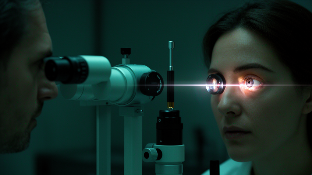
Every eye surgeon knows the frustration of a patient who insists their vision is failing while every standard test comes back normal. Visual acuity charts and contrast tests, the workhorses of ophthalmic clinics, often miss the subtle degradation of image quality that early cataracts, unstable tear films, or scattered light inside the eye can produce. A technology that fires a tiny point of light into the eye and watches it bounce back twice is now offering clinicians an objective way to measure what patients have been trying to describe all along, and a new narrative review published in Pharmacia pulls together a decade of scattered evidence into a single, clinically usable picture.
The technique, known as double-pass assessment and commercially available as the Optical Quality Analysis System, traces its origins to a 1956 experiment by Flamant, who first proposed measuring the point spread function of the human eye by imaging a point source on the retina. The modern version works in two stages. A near-infrared beam at 780 nanometers enters the eye through a controlled artificial pupil and is focused onto the retina in the first pass. Light reflected from the retinal surface then travels back out through the same cornea, lens, and vitreous in a second pass, and a charge-coupled device camera records the resulting image. Because the light samples the eye’s optics twice, the recorded retinal image captures the combined blurring effects of optical aberrations and intraocular light scatter, something wavefront aberrometers that measure aberrations alone systematically miss.
From that double-pass image, clinicians derive three principal numbers. The objective scatter index, or OSI, compares the peripheral to the central light energy in the point spread function, quantifying forward scatter that veils the retinal image; young healthy eyes typically score close to or below 1.0, with values climbing steadily with age. The modulation transfer function cutoff frequency, expressed in cycles per degree, describes how well the eye preserves contrast at fine spatial detail, with healthy values around or above 30 cycles per degree. The Strehl ratio, ranging from 0 to 1, summarizes overall optical fidelity against a diffraction-limited ideal. Higher OSI and lower MTF cutoff values objectively track reduced visual acuity and contrast sensitivity, giving clinicians quantitative anchors for complaints that previously rested on subjective description alone.
The technology’s diagnostic credentials in cataract are striking. In a landmark comparison against psychophysical straylight measurement with the C-Quant device, an OSI cutoff of 1.18 discriminated healthy from cataractous eyes with 89 percent sensitivity and 100 percent specificity. Larger studies have shown OSI correlates with all major cataract morphologies graded on the Lens Opacities Classification System III, with cortical opacities driving scatter and nuclear cataracts degrading the MTF cutoff through increased higher-order aberrations. Posterior subcapsular cataracts, the most visually treacherous type, elevate both scatter and aberration measures simultaneously. Yet the review is careful to warn against reducing surgery decisions to a single number: while OSI values of 3.0 or higher signal significant impairment, patients with lower values but genuine visual complaints still benefit from cataract surgery, so the metrics must complement, not replace, clinical judgment.
Repeatability data underpin the clinical case. In 119 healthy subjects, intraclass correlation coefficients exceeded 0.88 with narrow limits of agreement of roughly plus or minus 0.23 for OSI, and realignment of the eye between sessions added no meaningful variability. Repeatability limits widen in heterogeneous populations that include post-refractive surgery eyes and cataract patients, but the gap between normal and pathological values remains far larger than the measurement noise, preserving the device’s discriminative power even in difficult clinical territory. The review also flags genuine technical caveats: the built-in optometer shows a hyperopic bias of about half a diopter in high myopia, multiple scattering within the fundus can shift parameter estimates, and the standardized 4.0-millimeter artificial pupil means measurements may not reflect how the eye performs in dim light when natural pupils dilate.
Perhaps the most consequential application lies in refractive surgery planning, where the choice between implantable collamer lenses and corneal laser procedures can hinge on subtle differences in optical quality. Baseline measurements vary sharply with the degree of myopia: mean OSI rises from about 0.50 in low myopia to 1.33 in super-high myopia, while MTF cutoff falls from nearly 40 to under 28 cycles per degree. Comparative studies consistently show that phakic lens implantation induces fewer higher-order aberrations than LASIK, PRK, or SMILE. In a contralateral-eye comparison followed for three years, postoperative coma, spherical aberration, and total higher-order aberrations were all significantly lower after implantable collamer lens surgery, with safety and efficacy indices favoring the lens approach.
Corneal procedures, by contrast, produce a transient optical penalty. After PRK and LASIK, OSI rose by factors of 1.48 and 1.57 respectively at three months, and femtosecond LASIK outperformed conventional LASIK and PRK at six months with lower OSI and higher MTF values. After SMILE, OSI climbed from 0.75 preoperatively to 1.09 at twenty days before settling to 0.82 at three months, and patients with lower preoperative scatter achieved better final optical quality. At one year, however, changes in MTF cutoff and OSI after implantable collamer lens implantation were not significantly different from those after SMILE, suggesting the procedures converge over time. The review proposes practical interpretive zones: an OSI at or below 1.0 is reassuring, values between 1.0 and 1.5 warrant repeat measurement after optimizing refraction and the ocular surface, and values above 1.5 in a young eye without visible cataract demand a search for tear film, lens, vitreous, or fundal causes before any laser is fired.
The tear film emerges as a critical and often overlooked modifier. Because it is a dynamic optical surface, a single static measurement can miss the fluctuating vision that dry eye patients describe. Dynamic OSI protocols, sampling every half second across the interblink interval, reveal progressive deterioration within five to ten seconds in eyes with short tear breakup time, while normal eyes remain stable. Patients with meibomian gland dysfunction show more pronounced optical degradation than those with aqueous-deficient dry eye, and researchers have described ladder, seesaw, and plateau temporal patterns that may help subtype the disease. These dynamic indices correlate with tear breakup time and corneal staining, and they offer an objective way to monitor treatment response and even detect preclinical tear film instability in patients whose symptoms otherwise outpace their clinical findings.
The review’s authors are candid about what stands between this technology and routine adoption: heterogeneous study designs, variable measurement protocols, and the absence of universally validated diagnostic thresholds, particularly in borderline cases where scatter is elevated without clear lens changes. Their proposed framework, integrating cataract assessment, refractive surgery candidate selection, and ocular surface evaluation into one interpretive scheme, is explicitly a starting point rather than a finished standard, and prospective validation studies are the stated priority. Still, the direction of travel is clear. As populations age and demand ever-sharper vision after surgery, an instrument that turns the vague complaint of glare and blur into a reproducible number, one that can flag the cataract a slit lamp misses, steer a high myope toward a lens instead of a laser, and expose the unstable tear film behind fluctuating vision, may prove to be one of the quiet revolutions in modern eye care.
Subject of Research: Double-pass optical quality assessment using the objective scatter index for cataract evaluation and refractive surgery planning
Article Title: Clinical applications of double-pass optical quality assessment in cataract and refractive surgery: A narrative review
Article References: Milkovska, M., & Mermeklieva, E. (2026). Clinical applications of double-pass optical quality assessment in cataract and refractive surgery: A narrative review. Pharmacia, 73, Article e194149. https://doi.org/10.3897/pharmacia.73.e194149
Image Credits: AI Generated
DOI: 10.3897/pharmacia.73.e194149
Keywords: double-pass technology, objective scatter index, cataract, refractive surgery, OQAS, modulation transfer function, Strehl ratio, implantable collamer lens, LASIK, SMILE, dry eye, tear film
Cite Scienmag News
Ophelia Keating. (October 15, 2026). A Beam of Light Twice Through the Eye: New Tool Sharpens Cataract and Laser Surgery Decisions. Scienmag. https://scienmag.com/a-beam-of-light-twice-through-the-eye-new-tool-sharpens-cataract-and-laser-surgery-decisions/
Ophelia Keating. "A Beam of Light Twice Through the Eye: New Tool Sharpens Cataract and Laser Surgery Decisions." Scienmag, 15 October 2026, https://scienmag.com/a-beam-of-light-twice-through-the-eye-new-tool-sharpens-cataract-and-laser-surgery-decisions/. Accessed 15 October 2026.
Ophelia Keating. "A Beam of Light Twice Through the Eye: New Tool Sharpens Cataract and Laser Surgery Decisions." Scienmag. October 15, 2026. https://scienmag.com/a-beam-of-light-twice-through-the-eye-new-tool-sharpens-cataract-and-laser-surgery-decisions/


