


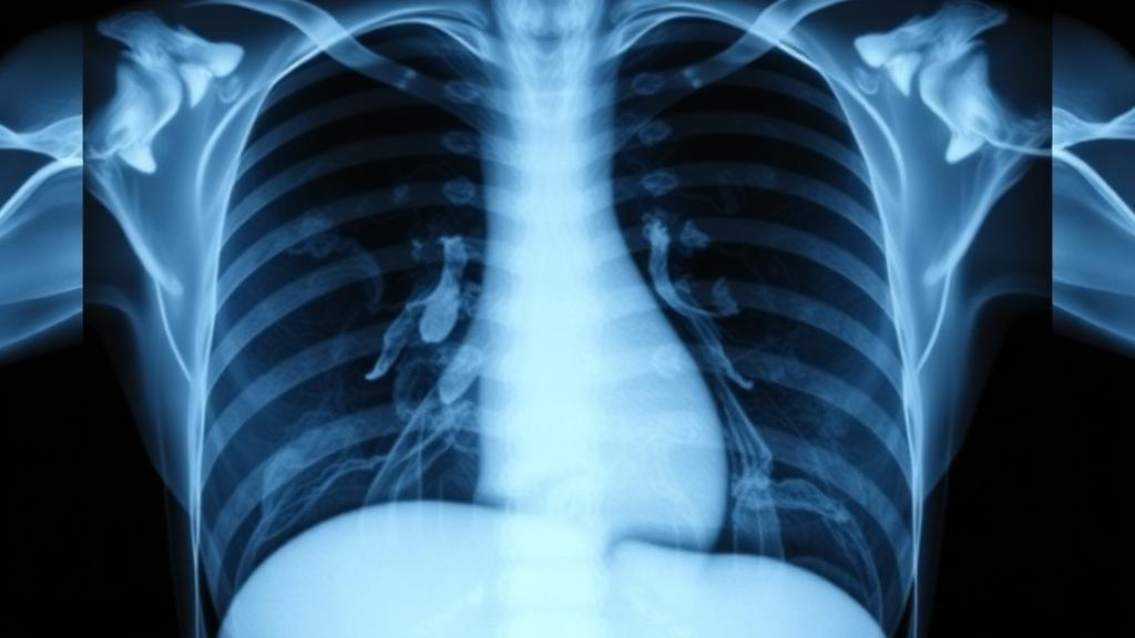
Radiologists around the world are drowning in images. Every chest X-ray that lands on a reading station must eventually be translated into a structured, clinically precise report, and that translation is still done largely by hand. A new study published in BMC Medical Imaging by Souha Aouadi and colleagues at Hamad Medical Corporation in Doha, Qatar, together with a collaborator at Hamad Bin Khalifa University, describes a comprehensive artificial intelligence workflow that automates this translation, converting paired chest radiographs into draft reports through a carefully engineered two-stage pipeline. The work arrives at a moment when imaging volumes are climbing faster than radiology workforces can grow, and it offers a technical blueprint that balances raw performance with something rarer in medical AI: interpretability.
The problem the researchers set out to address is deceptively simple to state and notoriously hard to solve. Writing a radiology report requires the radiologist to detect subtle findings in an image, organize them into clinically meaningful categories, and then express them in fluent, standardized prose. Manual report generation is time-consuming and error-prone, particularly for less experienced radiologists, and the resulting inefficiencies ripple through hospital workflows as case volumes increase. Automated radiology report generation, often abbreviated RRG in the machine learning literature, has therefore become one of the most active frontiers at the intersection of computer vision and natural language processing. The Qatar-based team argues that the most practical route to reliable automation is not a single monolithic model that maps pixels directly to paragraphs, but a decomposed workflow in which each stage can be inspected, evaluated, and improved independently.
Their pipeline begins with images and ends with text, but the crucial innovation lies in what happens in between. In the first stage, three multilabel classifiers built on the ConvNeXT deep learning architecture examine paired posterior-anterior and lateral chest X-rays and predict clusters of clinically relevant keywords. These keywords are not arbitrary; they are extracted from ground-truth radiology reports using KeyBERT, a keyword extraction model built on BERT-style language representations, and they serve as the target labels during training. In the second stage, a T5 model, short for Text-to-Text Transfer Transformer, takes the predicted keywords for each patient and generates a full radiology report from them. By forcing the system to pass through an intermediate keyword representation, the designers create a transparent bridge between what the image shows and what the report says, making it possible for a human reviewer to see exactly which visual findings the model detected before any prose was written.
The most distinctive technical contribution of the study is a frequency-aware clustering strategy for handling the keywords themselves. In medical datasets, the distribution of findings follows a steep power law: common terms such as those describing normal heart size or clear lungs appear thousands of times, while rare but clinically critical findings may appear only a handful of times. Previous keyword-based pipelines treated all labels uniformly, which meant that rare findings were effectively invisible to the learning algorithm. The proposed method instead partitions keywords into logarithmic frequency groups, specifically the ranges of ten to one hundred occurrences, one hundred to one thousand, and one thousand to ten thousand, and trains a dedicated classifier for each cluster. This design reduces label sparsity and improves the modeling of low-frequency findings, ensuring that the uncommon abnormalities that matter most in clinical practice are not drowned out by the vocabulary of routine normality.
Class imbalance is a persistent enemy in medical machine learning, and the authors attacked it with a second weapon beyond clustering. They employed binary cross-entropy-based focal loss in combination with transfer learning. Focal loss, originally introduced for dense object detection, down-weights the contribution of easy, well-classified examples so that gradient effort concentrates on the hard, ambiguous cases near the decision boundary. Transfer learning, meanwhile, allows the ConvNeXT backbones to start from visual representations learned on large natural image datasets rather than from scratch, a significant advantage when labeled medical data is scarce. Together, these techniques let the classifiers squeeze reliable signal out of a dataset that, while respectable in size, cannot match the millions of labeled images available in general-purpose vision benchmarks.
The evaluation was conducted on the publicly available IU X-ray dataset, also known as the Indiana University chest X-ray collection, which comprises 3,851 patients with paired posterior-anterior and lateral chest radiographs and their corresponding reports. The team divided the data into training, validation, and test sets using an 80 percent, 10 percent, 10 percent split, a standard configuration that allows fair comparison with prior work validated on the same benchmark. Because the dataset is public and widely used, the reported numbers can be placed directly alongside those of competing state-of-the-art systems, giving the community a clear yardstick for progress.
That yardstick tells a encouraging story. On the test set, the pipeline achieved a BLEU-1 score of 0.50, which the authors note represents an approximately 11 percent relative improvement over R2Gen, a widely cited baseline model for radiology report generation. The remaining BLEU scores, which measure n-gram overlap between generated and reference text at increasing levels of strictness, came in at 0.33 for BLEU-2, 0.22 for BLEU-3, and 0.16 for BLEU-4. The system also recorded a ROUGE-L score of 0.41, capturing the longest common subsequences between generated and reference reports, a METEOR score of 0.21, which accounts for synonymy and stemming in the comparison, and a CIDEr score of 0.37, a consensus-based metric that rewards descriptions agreeing with the judgments of multiple reference documents. Taken together, these figures indicate strong agreement between the generated reports and the human-written references, placing the keyword-mediated approach at or near the front of the pack for this benchmark.
What makes these numbers more than a leaderboard entry is the interpretability argument that underpins the entire design. End-to-end models that generate reports directly from images are famously opaque: when a generated report contains an error, clinicians have no principled way to determine whether the failure occurred in visual perception, in clinical reasoning, or in language generation. The two-stage architecture localizes the decision points. A reviewer can inspect the predicted keywords and immediately judge whether the model saw what a radiologist would see, before worrying about how elegantly the T5 model phrases the finding. This design enhances transparency by linking image features to clinically relevant terms prior to report generation, and it also means the keyword classifiers can be audited with familiar classification metrics such as accuracy, precision, recall, F1-score, and area under the ROC curve, tools that regulators and hospital quality teams already understand.
The study, supported by the Medical Research Center at Hamad Medical Corporation under study number MRC-03-25-1173, was published as open access under a Creative Commons license, and the authors received the article’s acceptance after review in the spring of 2026. The team is candid about the road ahead. Their future work will focus on extending the approach to three-dimensional imaging modalities, a substantial challenge because computed tomography and magnetic resonance volumes contain far richer spatial structure than two-dimensional radiographs, and on validating performance on clinical datasets drawn from real hospital environments rather than curated public benchmarks. Such validation will be essential before any automated drafting system can sit credibly inside a radiologist’s workflow.
Even so, the trajectory is clear and, for a profession under mounting pressure, welcome. The Doha team has shown that a decomposed, frequency-aware, keyword-mediated pipeline can outperform direct image-to-text generation on a standard benchmark while giving clinicians a window into the machine’s reasoning. If the approach survives contact with three-dimensional data and live clinical deployments, the routine chest X-ray report of the future may well be drafted by an algorithm in seconds and refined by a human in minutes, a division of labor that could return the most scarce resource in radiology, attentive expert time, to the cases that need it most.
Subject of Research: Automated two-stage AI generation of radiology reports from chest X-ray images
Article Title: Comprehensive AI workflow for radiological report generation from radiographs
Article References: Comprehensive AI workflow for radiological report generation from radiographs. (n.d.). https://doi.org/10.1186/s12880-026-02464-3
Image Credits: AI Generated
DOI: 10.1186/s12880-026-02464-3
Keywords: radiology report generation, chest X-ray, deep learning, ConvNeXT, T5 transformer, KeyBERT, multilabel classification, focal loss, IU X-ray dataset, BLEU, interpretability, medical imaging AI
Cite Scienmag News
Ophelia Keating. (October 15, 2026). AI Turns Chest X-rays into Draft Radiology Reports with a Two-Stage Keyword Pipeline. Scienmag. https://scienmag.com/ai-turns-chest-x-rays-into-draft-radiology-reports-with-a-two-stage-keyword-pipeline/
Ophelia Keating. "AI Turns Chest X-rays into Draft Radiology Reports with a Two-Stage Keyword Pipeline." Scienmag, 15 October 2026, https://scienmag.com/ai-turns-chest-x-rays-into-draft-radiology-reports-with-a-two-stage-keyword-pipeline/. Accessed 15 October 2026.
Ophelia Keating. "AI Turns Chest X-rays into Draft Radiology Reports with a Two-Stage Keyword Pipeline." Scienmag. October 15, 2026. https://scienmag.com/ai-turns-chest-x-rays-into-draft-radiology-reports-with-a-two-stage-keyword-pipeline/


