


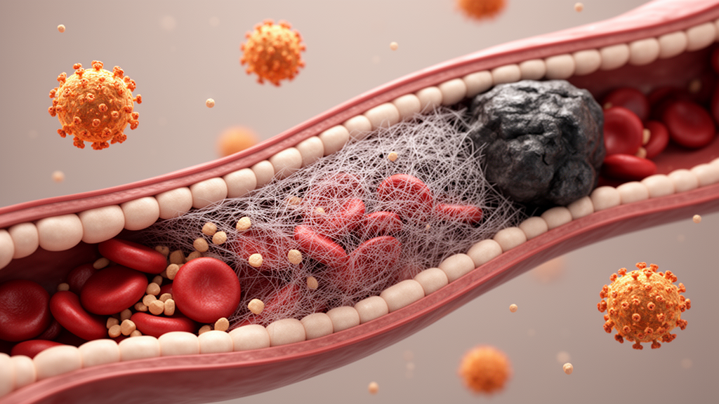
Dengue fever has long been defined by its dangers of bleeding: leaking plasma, crashing platelets, and hemorrhagic shock dominate the textbooks and the public imagination alike. But a newly published case report in Clinical Case Reports serves as a striking reminder that the mosquito-borne virus can push the body’s clotting machinery in the opposite direction, with potentially fatal consequences. Physicians describe a 25-year-old man who, while apparently recovering from dengue, developed deep vein thrombosis in his right leg and bilateral pulmonary emboli that strained his heart. The case, published open access by a team led by Sarah Nisar and colleagues, adds to a small but growing body of evidence that venous thromboembolism is a rare yet real complication of dengue, one that clinicians may easily miss precisely because their attention is fixed on the bleeding risk.
The patient arrived at the emergency department with three days of high-grade fever, reaching 103 degrees Fahrenheit, accompanied by the classic hallmarks of dengue: headache, retro-orbital pain behind the eyes, and generalized body aches. He had no vomiting, diarrhea, cough, or any bleeding manifestations, and no chronic medical conditions, prior surgeries, or relevant family history. He did not smoke, drink alcohol, or use recreational drugs. On examination he appeared ill and febrile, with a pulse of 110 beats per minute and a blood pressure of 115/80 mmHg, but his heart, lungs, abdomen, and neurological status were otherwise unremarkable. In short, he presented as a textbook case of uncomplicated dengue.
Laboratory testing confirmed the diagnosis. The dengue NS1 antigen came back positive, malaria parasites were absent, and ELISA-based serology ruled out other viral infections. His hemoglobin stood at 13 g/dL and his platelet count at 192,000 per microliter, well within the normal range, which would later prove to be one of the most instructive features of the case. His leukocyte count was initially elevated at 12,000 per microliter before settling to 4,600. More ominous was the coagulation picture: while his standard coagulation profile was normal, his D-dimer, a breakdown product of cross-linked fibrin that serves as a molecular flag for active clot formation and dissolution, was dramatically elevated at 9,968 ng/mL, later declining to 4,068 ng/mL. Inflammatory markers told a similar story of a body under siege, with C-reactive protein at 142 mg/L against a normal threshold of 10, procalcitonin at 0.57 ng/mL, and an erythrocyte sedimentation rate of 54 mm/h.
The turning point came on the sixth day of hospitalization, roughly the ninth day of illness, when the patient developed sudden painful swelling of the right lower limb extending from calf to thigh, together with progressive shortness of breath on exertion. The limb was visibly swollen up to the groin, red, warm, and tender, with increased calf and thigh circumference, though peripheral pulses remained intact. He remained hemodynamically stable apart from mild tachycardia, with a blood pressure of 105/70 mmHg and an oxygen saturation of 93 percent on room air. Doppler ultrasonography confirmed deep vein thrombosis, and because his dyspnea worsened, the team proceeded to CT pulmonary angiography. The scan revealed bilateral pulmonary emboli and dilatation of the right cardiac chambers, a sign that the clots lodging in the pulmonary arteries were forcing the right side of the heart to pump against dangerous resistance.
Imaging then uncovered a further puzzle. A chest radiograph showed a cavitary lesion in the right lung with mild cardiomegaly, and high-resolution computed tomography revealed that cavity surrounded by pneumonic consolidation, minimal bilateral pleural effusions, and basal atelectatic changes. Cavitary lung lesions typically raise the specter of tuberculosis, lung abscess, fungal infection, or malignancy, each of which could independently predispose a patient to clotting. The team therefore cast a wide diagnostic net: sputum examination, acid-fast bacilli smears, and GeneXpert testing were negative for Mycobacterium tuberculosis, blood cultures grew no bacterial or fungal pathogens, and bronchoscopy with bronchoalveolar lavage found only mucopurulent secretions with no organisms on Gram stain, no growth on culture, and no malignant cells on cytology. Transthoracic echocardiography showed preserved left ventricular function with an ejection fraction of 64 percent, no intracardiac vegetations, and a positive McConnell sign, a regional pattern of right ventricular dysfunction considered suggestive of acute pulmonary embolism.
The authors argue that the most likely explanation for the cavity is pulmonary infarction, tissue death downstream of the embolic obstruction, which can occasionally cavitate when vascular blockage is severe. The temporal relationship between the dengue infection and the thrombotic event, combined with a negative thrombophilia workup and the patient’s young age, pointed toward a transient, infection-associated prothrombotic state rather than an inherited clotting disorder or occult cancer. The differential diagnosis nonetheless included septic pulmonary emboli, secondary bacterial pneumonia, and malignancy, and the authors note that unexplained or recurrent venous thromboembolism should always prompt consideration of hidden malignancy, citing a prior report in which deep vein thrombosis was the presenting feature of uterine leiomyosarcoma.
The mechanistic picture emerging from the dengue literature is one of coordinated disruption of hemostasis. The authors outline several converging pathways: molecular mimicry between viral antigens and coagulation factors generating cross-reactive autoantibodies; direct endothelial dysfunction driven in part by the dengue NS1 protein; infection-induced upregulation of tissue factor in monocytes and endothelial cells; and a cytokine storm that amplifies both inflammation and coagulation activation. Critically, this case demonstrates that clinically significant thrombosis can occur even when platelet counts remain normal. Previous analyses have documented venous thrombosis in dengue patients with median platelet counts as low as 52,000 per cubic millimeter, but this patient never developed thrombocytopenia during hospitalization, underscoring that the absence of low platelets offers no reassurance against the thrombotic complication.
The timing also fits an emerging pattern. In an analysis of 19 previously reported cases of spontaneous venous thrombosis in dengue cited by the authors, the median onset of thrombosis was six days after fever began, with serious thrombotic events generally clustering between days 5 and 10 of illness, the transition from the acute phase to recovery. Lower limb deep vein thrombosis was the dominant manifestation, accounting for 68 percent of cases, most often involving the right side and the external iliac, common femoral, and popliteal veins. This patient’s right lower limb clot extending from calf to thigh, with bilateral pulmonary embolism on top, represents an unusually severe expression of that same tendency. His markedly elevated C-reactive protein also aligned with evidence that more intense inflammatory responses accompany severe dengue, where mean CRP levels of roughly 50 mg/L have been recorded against about 22 mg/L in non-severe disease.
Treatment required navigating a genuine therapeutic dilemma: anticoagulating a dengue patient in whom bleeding is the classically feared complication. The team initiated weight-based therapeutic enoxaparin at 60 mg subcutaneously twice daily for the approximately 60-kg patient, alongside intravenous paracetamol, fluids, broad-spectrum antimicrobials with meropenem and linezolid given concern for secondary bacterial infection, and nebulized bronchodilators and corticosteroids for respiratory symptoms. In the published series, 57.8 percent of patients received low-molecular-weight heparin, anticoagulation was tolerated even amid thrombocytopenia, and all patients recovered without incident. Yet the literature still lacks formal guidelines for managing dengue-associated pulmonary embolism, and the authors emphasize that the decision to anticoagulate remains difficult in the face of bleeding risk. This patient improved steadily, was discharged on day thirteen with apixaban 10 mg twice daily tapering to 5 mg twice daily for a total of six months of anticoagulation, and at four-week follow-up his dyspnea and leg swelling had resolved, repeat Doppler ultrasound showed interval improvement, and chest CT showed partial resolution of the cavitary lesion.
The case carries two forward-looking warnings. First, because the patient demonstrated right ventricular dysfunction from bilateral pulmonary emboli, he faces a risk of chronic thromboembolic pulmonary hypertension, a disabling late complication that demands extended monitoring. Second, and more broadly, the authors urge clinicians caring for dengue patients to stay alert for thrombotic symptoms during the recovery phase, when vague complaints such as unilateral limb swelling or exertional breathlessness may be the only clues to a clot that imaging will confirm. Early recognition, timely diagnosis with tools such as D-dimer measurement, Doppler ultrasound, and CT angiography, and prompt anticoagulation can prevent life-threatening outcomes in a complication that remains poorly recognized but is, as this case shows, very much preventable.
Subject of Research: Venous thromboembolism as a complication of dengue fever
Article Title: Post‐Dengue Deep Vein Thrombosis Complicated by Pulmonary Embolism: A Case Report
Article References: Nisar, S., Mushtaq, H., Khan, M. H., Sajjad, F., Afridi, A., Fatima, N. E., & Kamil, K. A. (2026). Post‐Dengue Deep Vein Thrombosis Complicated by Pulmonary Embolism: A Case Report. Clinical Case Reports, 14(10), Article e73642. https://doi.org/10.1002/ccr3.73642
Image Credits: AI Generated
DOI: 10.1002/ccr3.73642
Keywords: dengue, deep vein thrombosis, pulmonary embolism, venous thromboembolism, hypercoagulability, D-dimer, anticoagulation, enoxaparin, apixaban, NS1 antigen, pulmonary infarction, case report
Cite Scienmag News
Ophelia Keating. (October 9, 2026). Dengue Recovery Hides a Deadly Clot Risk, Case Report Warns. Scienmag. https://scienmag.com/dengue-recovery-hides-a-deadly-clot-risk-case-report-warns/
Ophelia Keating. "Dengue Recovery Hides a Deadly Clot Risk, Case Report Warns." Scienmag, 9 October 2026, https://scienmag.com/dengue-recovery-hides-a-deadly-clot-risk-case-report-warns/. Accessed 9 October 2026.
Ophelia Keating. "Dengue Recovery Hides a Deadly Clot Risk, Case Report Warns." Scienmag. October 9, 2026. https://scienmag.com/dengue-recovery-hides-a-deadly-clot-risk-case-report-warns/


